A clear view
Dr. Peter Kardos' eyes became increasingly cloudy. Against the light, he could only see contours; he could no longer recognize curves on the monitor. The reason was a dysfunction of the endothelial cells. His cornea swelled up. On recommendation, he underwent treatment at Freiburg University Hospital. Following a minimally invasive procedure, in which Medical Director Prof. Thomas Reinhard transplanted only the inner layer of the cornea rather than the entire cornea, he can now see clearly again.
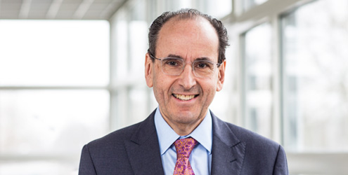
Dr. Peter Kardos likes to walk the few meters from his home to his joint practice. During this walk, the doctor noticed for the first time that something was wrong with his eyes. The morning sun was shining towards him and he couldn't clearly make out the faces of people walking towards him. He could only see outlines. His ophthalmologist initially diagnosed a cataract, but it was still at too early a stage to be the cause of his vision problems. The reason for the increasing clouding of his cornea was what is known as Fuchs' corneal endothelial dystrophy - a congenital disease in which the endothelial cells that separate the cornea from the inside of the eye are no longer able to pump fluid out of the cornea with increasing age. The cornea swells. Visual acuity decreases and sensitivity to glare increases. "I saw as if through a fog. In the sleep lab, I could no longer make out some of the colored curves on the monitors," recalls Dr. Peter Kardos.
First, the lens affected by the cataract was removed and a new one implanted. The procedure was also intended to protect the cornea. However, the clouding then worsened. Dr. Kardos: "I could see even worse than before the operation. My ophthalmologist then recommended treatment at the eye clinic at Freiburg University Hospital, where a gentle procedure could be used to treat my corneal opacity."
Suture-free transplantation
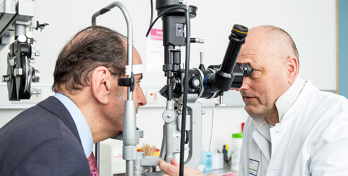
The endothelium is not capable of regeneration, so for a long time a corneal transplant was the only way to restore its function. The Eye Clinic at the University Hospital is increasingly using a gentler, minimally invasive method, Descemet's Membrane Endothelial Keratoplasty (DMEK). This involves replacing only the affected inner layer of the cornea rather than the entire cornea. During the procedure, the diseased layer is first removed via a small incision and then the transplant is fixed in the eye without sutures using an air bubble.
Dr. Kardos was admitted to the University Hospital as an inpatient. "I was amazed at how well organized the eye clinic is," says the Frankfurt native. "The operation took an hour. My left eye was operated on first, then my right eye a few months later." He had to lie on his back for three to four days afterwards so that the implanted cell layer would bond with the cornea. The first operation went without complications. However, in 30 percent of cases, the cell layer does not attach immediately, so that new air has to be filled in. This was also the case after the operation on Dr. Kardos' right eye. "However, the second operation was successful and I can now see very well in both eyes." In future, he will be able to sit comfortably in the back row at lectures again and still recognize every word in the presentations, he adds happily.
Corporate Communications
Breisacher Straße 153
79110 Freiburg
Phone: 0761 270-84830
Fax: 0761 270-9619030
kommunikation@uniklinik-freiburg.de