Understanding brain diseases better with highlighters
Thanks to a novel method, important types of immune cells in the brain can be differentiated for the first time and their role in brain diseases can be specifically investigated / Study published in the journal Nature Immunology
Parkinson's, Alzheimer's, multiple sclerosis: immune cells of the brain are probably involved in all neurological diseases. However, the role of different cell types has so far been difficult to assess. Now, an international team of researchers led by the Medical Center - University of Freiburg, together with scientists from Charité - Universitätsmedizin Berlin, the Technical University of Munich and the Weizmann Institute of Science in Israel, has gained new insights into the brain's own immune system. Using the novel, high-resolution technique of single-cell analysis, the researchers were able to distinguish individual types of the brain's own immune cells, known as microglia, and examine them separately from immune phagocytes, which only migrate into the brain in the course of a person's life. To do this, the interdisciplinary team first identified genetic characteristics that only occur in the target cells and then switched off the cells with the help of genetic tools - or made them glow. In this way, the specific contribution of different immune cells in the brain can now be investigated. The method, which was published in the journal Nature Immunology on June 15, 2020, can now be used by researchers worldwide.
"Until now, we always had to look at different types of immune cells in the brain at the same time. With our new approach, we can now for the first time precisely investigate the role of different immune cells in the brain in neuronal diseases," says project leader Prof. Dr. Marco Prinz, Medical Director of the Institute of Neuropathology at the Medical Center - University of Freiburg and member of the Freiburg Cluster of Excellence CIBSS - Centre for Integrative Biological Signalling Studies at the University of Freiburg.
Exploring the brain's own immune system with molecular genetics and luminescent proteins
Using the CRISPR/Cas9 system, a so-called gene scissors, the researchers were able to change DNA building blocks in the genetic material of mice depending on the cell type so that microglial cells were color-coded or switched off. By coloring the immune cells, researchers will also be able to gain new insights into communication with nerve cells in the future.
A major problem for neuroscientists to date has been that, in addition to microglial cells in the depths of the brain, there are also macrophages on the surface of the brain and around the blood vessels, for which different functions have been assumed but which have not yet been demonstrated. Scientists worldwide can now use the methods developed in Freiburg to specifically influence microglia in mice and thus investigate the role of macrophages in more detail. "The diversity of macrophages and their targeted modification will open up new and more specific therapeutic approaches in the future," summarizes Prinz.
As the brain's own immune guardians, microglia perform many different tasks in the healthy and diseased brain, from supplying nutrients to repairing tissue. In recent years, they have increasingly been attributed an important role in the development of numerous degenerative brain diseases such as Alzheimer's and Parkinson's, but also in inflammatory diseases such as multiple sclerosis and brain tumors. The majority of genetic risk factors for Alzheimer's and multiple sclerosis are found in microglial cells in the brain. Scientists around the world are therefore very interested in understanding microglial cells in more detail in order to be able to modify them therapeutically in the future.
" Microglial cells are also crucial for brain development," says Lukas Amann, biologist and one of the first authors from the Institute of Neuropathology at the Medical Center - University of Freiburg. "Since they cap excess nerve cell connections in the child's brain, microglia are also seen as potential target cells for therapeutics in developmental neuropsychiatric diseases," says Amann. As these can now be examined more closely in animal models using the novel methods developed by the Freiburg researchers, a much more precise cellular picture of the diseased brain should soon emerge.
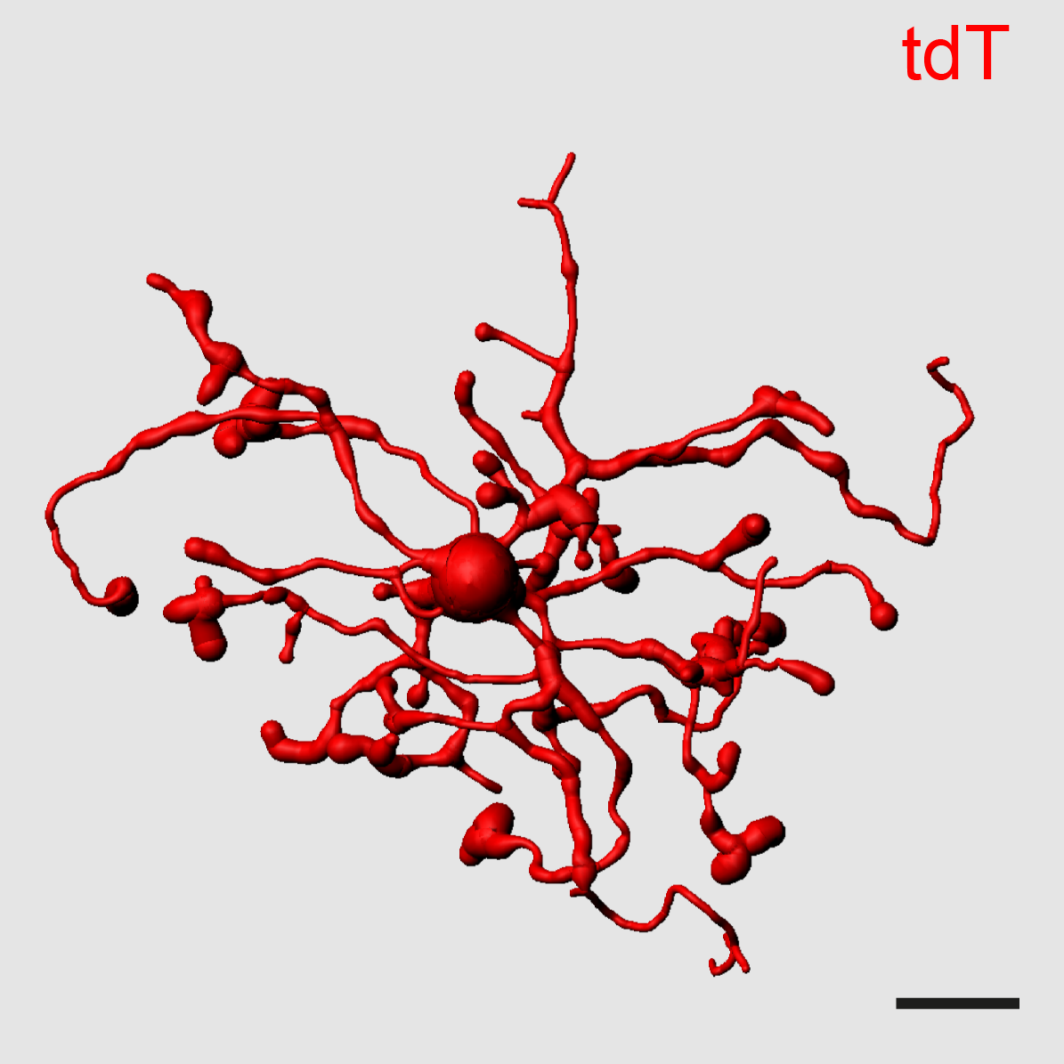
Image: Three-dimensional representation of a microglial cell in the brain. With the new imaging method, these immune guardians can be visualized and manipulated in detail with all their extensions in the healthy and diseased brain in high resolution.
Image source: Medical Center - University of Freiburg/Lukas Amann
Original title of the study: Novel Hexb-based tools for studying microglia in the CNS
DOI: 10.1038/s41590-020-0707-4
Link to the study: https://www.nature.com/articles/s41590-020-0707-4
Contact:
Prof. Dr. Marco Prinz
Institute of Neuropathology
Uniklinik Freiburg
Phone: 0761 270-51060
marco.prinz@uniklinik-freiburg.de
Back
Medical Center - University of Freiburg
Central Information
Phone: 0761 270-0
info@uniklinik-freiburg.de
For press inquiries:
Corporate Communications
Breisacher Straße 153
79110 Freiburg
Phone: 0761 270-84830
kommunikation@uniklinik-freiburg.de