Immune guardians in the human brain remeasured
Various manifestations of immune cells in the human brain identified for the first time / Special subtype of immune guardians discovered in brain tumors / Study published in the journal Nature Neuroscience
An international team of researchers led by the University Medical Center Freiburg, the Max Planck Institute of Immunobiology and Epigenetics Freiburg and Charité - Universitätsmedizin Berlin has remeasured the human brain's own immune system in healthy and diseased brains. The researchers found a surprising number of different manifestations of immune cells called microglia. Using novel, high-resolution techniques, the team from Freiburg and Berlin investigated the building blocks and metabolism of individual immune cells in brain tissue. In this way, they demonstrated in detail how the human immune system changes in brain tumors, which could be important for future therapeutic approaches. The study was published on November 18, 2019 in the journal Nature Neuroscience.
"We were very surprised to see how many different forms microglial cells can be found in the human brain. The state of the cells is obviously strongly influenced by factors such as ageing, tumor activity and surrounding cells," says project leader Prof. Dr. Marco Prinz, Medical Director of the Institute of Neuropathology at the Freiburg University Medical Center and member of the Cluster of Excellence CIBSS - Centre for Integrative Biological Signalling Studies at the University of Freiburg. "The diversity of immune cells opens up new therapeutic approaches against brain tumors or neurodegenerative diseases," summarizes Prinz. For their study, the researchers analyzed tissue samples from 15 patients from whom brain tissue had to be removed due to epilepsy or a tumor. Previous studies on rodent brains had come to the conclusion that microglia can only assume a few different activity states.
Microglia, the immune guardians in the brain, take on many different tasks during brain development as well as in healthy and diseased adult brains, from nutrition to tissue repair. In recent years, these brain's own immune guardians have increasingly been attributed an important role in the development of numerous degenerative brain diseases such as Alzheimer's and Parkinson's, but also in inflammatory diseases such as multiple sclerosis and brain tumors. Scientists around the world are therefore very interested in understanding microglial cells in more detail in order to be able to modify them therapeutically in the future.
A ring full of different immune guardians in brain tumors
The researchers led by Prinz and Dr.Dominic Grün, research group leader at the Max Planck Institute of Immunobiology and Epigenetics in Freiburg, together with the first authors of the study, Dr. Roman Sankowski from the Institute of Neuropathology at the University Medical Center Freiburg, and Dr. Chotima Böttcher from Charitè University Medicine Berlin, compared the different states of microglial cells in human brain tumors in detail. Until now, it was assumed that it is primarily immune cells circulating in the blood that are found in brain tumors. Sankowski was able to show that there are specially activated brain microglial cells in brain tumor tissue. These cells differ from other microglia in terms of their cellular make-up and cell metabolism. "We hope that new, more cell-specific therapeutic approaches with fewer side effects can now be developed to better treat tumor diseases," says Sankowski.
Exploring the cell with laser and molecular analysis
The study was made possible thanks to newly developed single cell analyses. The researchers used RNA analyses to determine gene activity and laser measurements to determine the protein content of individual cells extracted from the brain tissue. "These methods give us a much more precise cellular picture of very complex tissues such as the brain and the changes taking place in them," says Grün, one of the developers of this technology. "The methods should therefore have enormous potential for medical diagnostics," says Grün.
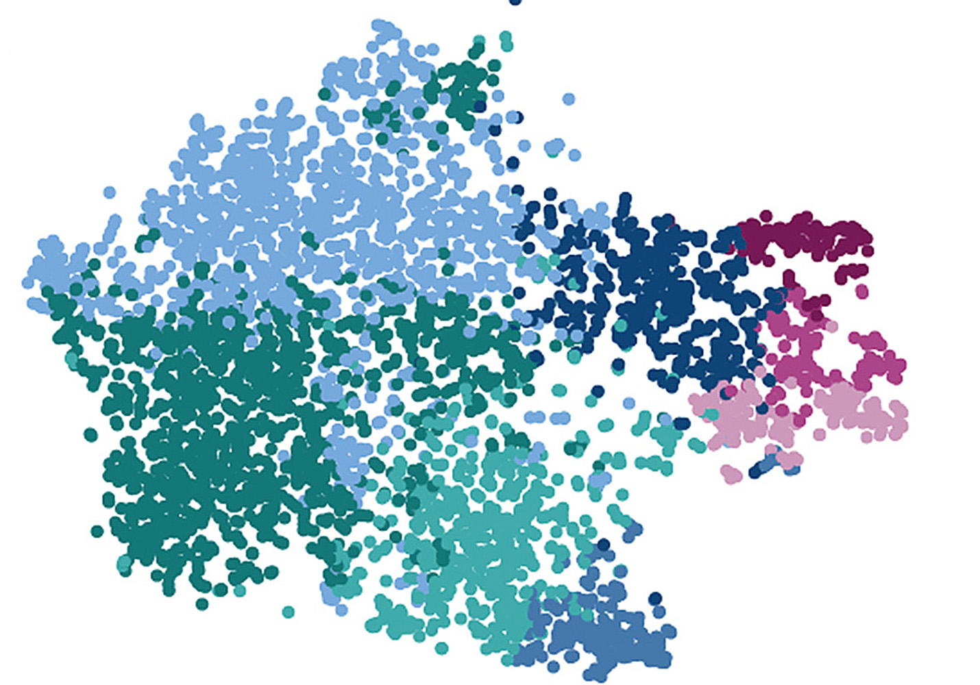
Caption: Single cell analysis of microglial cells: Each dot shows a cell and the colors signal different groups of microglial cells as they occur in the human brain.
Copyright: Roman Sankowski / University Medical Center Freiburg
Original title of the study: Mapping microglia states in the human brain through the integration of high-dimensional techniques.
DOI: 10.1038/s41593-019-0532-y
Link to the study: www.nature.com/articles/s41593-019-0532-y
Contact:
Prof. Dr. Marco Prinz
Institute of Neuropathology
Freiburg University Medical Center
Phone: 0761 270-51060
marco.prinz@uniklinik-freiburg.de
Dr. Dominic Grün
Max Planck Institute of Immunobiology and Epigenetics, Freiburg
Stübeweg 51
Phone: 0761 5108490
gruen@ie-freiburg.mpg.de
Back
Medical Center - University of Freiburg
Central Information
Phone: 0761 270-0
info@uniklinik-freiburg.de
For press inquiries:
Corporate Communications
Breisacher Straße 153
79110 Freiburg
Phone: 0761 270-84830
kommunikation@uniklinik-freiburg.de