Fast MRI quantum technology for tumor diagnostics
New imaging method with hyperpolarized contrast agent allows imaging of tumour metabolism / Use of quantum technology should significantly simplify the procedure / First patient studies on prostate cancer planned
The Federal Ministry of Education and Research (BMBF) is now funding an innovative project to improve the imaging of tumors with 15.8 million euros over four years. Of this, 3.1 million euros will go to the Medical Center - University of Freiburg as a clinical partner. The structure of tumors can be depicted very well in diagnostic magnetic resonance imaging (MRI). However, decisive information on tumor metabolism has been lacking to date. The so-called hyperpolarization method makes it possible to visualize this. In the project now being funded, the aim is to use quantum technology to make this previously promising but very complex technique much faster and cheaper and soon make the step into clinical care.
"We hope that cancer patients will benefit from imaging of tumour metabolism using hyperpolarization MRI as soon as possible. Our preliminary methodological work in recent years has laid an important foundation for this," says Prof. Dr. Michael Bock, physicist and Professor of Experimental Radiology at the Department of Radiology at the Medical Center - University of Freiburg. "In the project that is now starting, we will be able to test the enormous potential of this technology for the tumor diagnostics of tomorrow on a broad scale for the first time," says Dr. Andreas Schmidt, Group Leader in the Department of Radiology - Department of Medical Physics.
Enormous potential of the hyperpolarization method known from studies
Tumor metabolism can be investigated and even quantified using special MRI procedures based on the body's own substance pyruvate, for example. To do this, the substance must be prepared in such a way that its weak MRI signal is amplified by a factor of 100,000 or more. Until now, this so-called hyperpolarization took several hours and was technically very complex, so that clinical use could only be investigated in a few studies - but these studies have already shown the great potential for oncology.
As part of the "Quantum technologies - from the basics to the market" programme, the BMBF is funding a new quantum-based technology with which hyperpolarized MRI contrast agents can be produced in just a few minutes directly before the examination at significantly lower costs. The collaborative project "QuE-MRT: Revolutionizing cancer imaging through quantum technologies" combines the experience of the external project partner, the company NVision in Ulm, which produces the novel hyperpolarizer, with the clinical and methodological expertise at the university hospitals in Freiburg, Munich and Ulm. All studies are coordinated by the Clinical Trials Unit at Freiburg University Hospital.
Focus on prostate tumors
In order to optimize the new technology for use in clinical routine and subsequently test it on patients, each hospital site focuses on different types of tumors. The Medical Center - University of Freiburg is concentrating on prostate cancer in its sub-study. "The altered metabolism is an important indicator that can be used to distinguish aggressive prostate carcinomas from less aggressive ones," says Prof. Dr. Christian Gratzke, Medical Director of the Department - University of Freiburg's Department of Urology. MRI already plays a significant role in the diagnosis of patients with prostate cancer. "With hyperpolarized contrast agents, we could significantly expand the diagnostic spectrum and thus adapt cancer therapy even more individually in the future," says Prof. Dr. Fabian Bamberg, Medical Director of the Department of Radiology at the Medical Center - University of Freiburg.
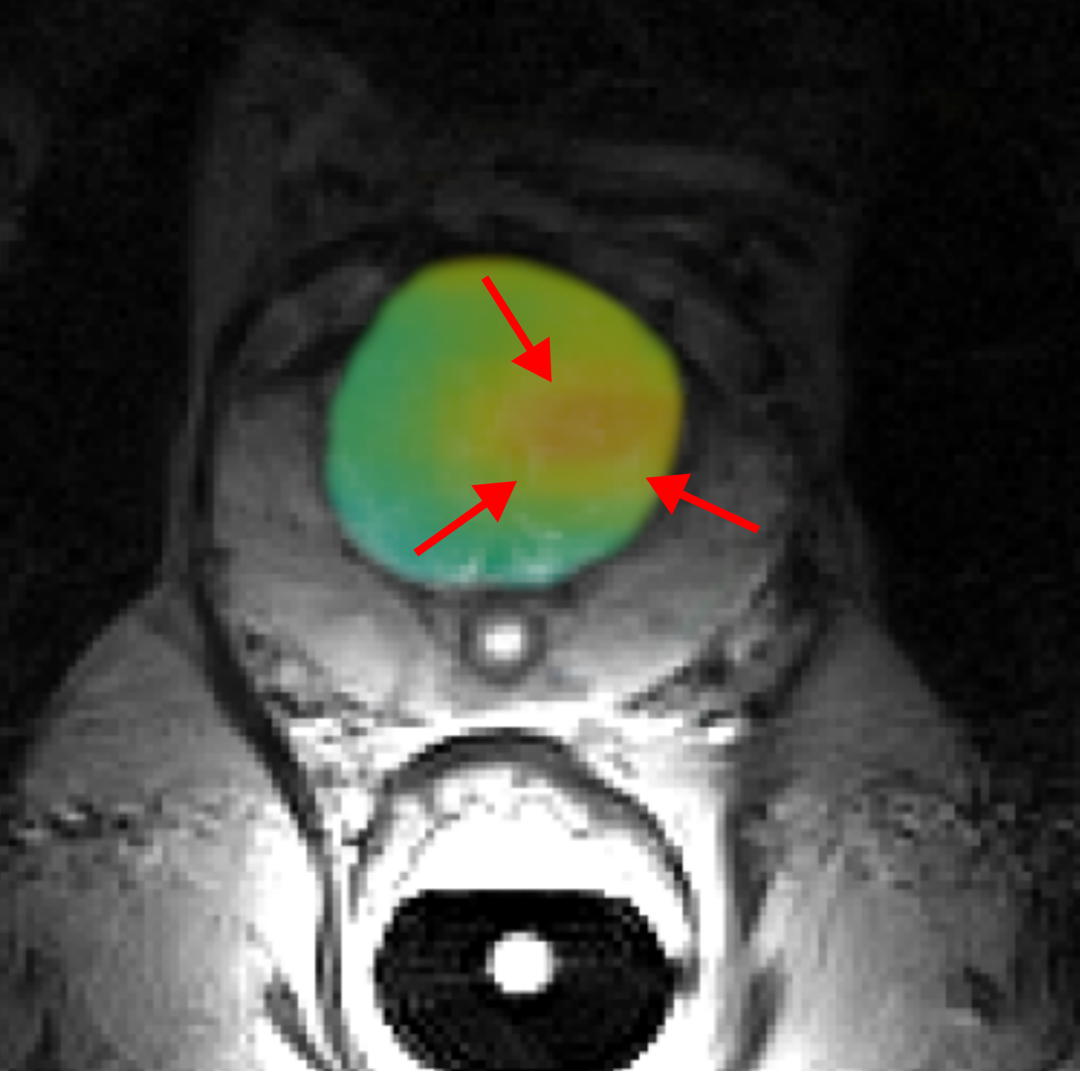
Caption: In the conventional MRI image (grey), the organs in the abdominal cavity can be seen. In the area of the prostate, the metabolism of the pyruvate molecule is shown in colour, the signal of which was amplified with the help of hyperpolarization. The metabolism is particularly high in the light red area, which indicates a tumor-like change.
Image source: C. A. Müller/University Center - University of Freiburg
Contact:
Prof. Dr. Michael Bock
Head of the Department of Experimental Radiology
Medicine Physics of
Department of Radiology
Medical Center - University of Freiburg
Phone: 0761 270-94140
michael.bock@uniklinik-freiburg.de
Back
Medical Center - University of Freiburg
Central Information
Phone: 0761 270-0
info@uniklinik-freiburg.de
For press inquiries:
Corporate Communications
Breisacher Straße 153
79110 Freiburg
Phone: 0761 270-84830
kommunikation@uniklinik-freiburg.de