Treatment of bile stasis using drainage (PTCD)
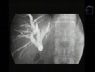
X-ray image of bile ducts after injection of contrast medium. A constriction in the common bile duct can be seen.
Bile outflow disorders can lead to a build-up of bile in the draining bile ducts. In most cases, this blockage can be removed during an endoscopy. However, after previous operations in the upper abdomen (e.g. stomach), it is sometimes not possible to reach the bile duct orifice in this way. An attempt is then made to remove the bile blockage by puncturing the bile ducts through the skin.
An obstructed bile duct in the liver is first searched for using ultrasound. After disinfecting the skin, a local anaesthetic is applied under sterile conditions to the skin and the liver capsule in the area of the right flank or upper abdomen, which borders the abdominal wall at this point. A thin needle is then inserted through the skin into the liver.
By carefully injecting small amounts of X-ray contrast medium and simultaneously advancing or withdrawing the needle under X-ray control, a bile duct is located. As soon as bile ducts are contrasted, a wire is inserted into the bile duct and a drainage tube (PTCD) is inserted over it, which drains the bile either outwards or inwards. The correct medical term for this is percutaneous transhepatic cholangiodrainage (PTCD). In rare cases, the liver, pleura or lungs may be injured during the examination and, in rare cases, bleeding from the puncture site or infections may occur.
Before the examination
The patient will be informed about the examination, which is only carried out as an inpatient, by the attending physician. Medication that inhibits blood clotting must be discontinued.