Experimental Neurorehabilitation Laboratory
Mission
The integrity of brain circuits is threatened by pathological conditions like vascular injury or (para-) infectious disease. Especially in the phase after the acute damage, neurorehabilitation is the only therapeutic approach to reduce dependency and disability in daily life. Our mission is to investigate pathophysiology of disease and mechanisms underlying the restoration of function. More specifically, our focus is on (1) research into the recovery of motor deficits after stroke, (2) research into fatigue and cognitive impairment after COVID-19, and (3) research into the functional significance of network integrity after stroke and neurodegenerative diseases. For this reason, we not only carry out basic research in the rodent model. The transferability of relevant mechanisms and findings is also examined in humans using the most modern MRI-based procedures.
Research topics
Topic 1: Role of dopaminergic signalling for motor learning and recovery after stroke:
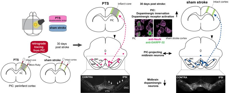
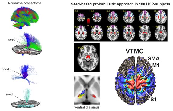
We identified and characterized dopaminergic projections from the ventral midbrain to the primary motor cortex in rats that are required for acquisition of novel movement sequences and motor learning [1], [2]. A homologue of this projection could also be identified in humans using a Diffusion-Tensor-Imaging-based global tractography approach [3]. After ischemic stroke, dopaminergic neurons within the ventral midbrain degenerate, even though the ischemic lesion is in a distant location (i.e. exo-focal degeneration) [4]. In patients that survived an ischemic stroke, degeneration of connections between ventral midbrain and M1 is linked to an adverse outcome regarding independence in daily life, upper limb motor function and non-motor symptoms [5]. Preventing degeneration of dopaminergic neurons using a neuroprotective agent (Substance P) also improved motor rehabilitation in a rat model of focal stroke [6]. Thus, neuroprotective approaches preventing exo-focal degeneration of dopaminergic neurons are an innovative and promising approach to support neurorehabilitation after stroke.
[1] J. A. Hosp, A. Pekanovic, M. S. Rioult-Pedotti, und A. R. Luft, „Dopaminergic projections from midbrain to primary motor cortex mediate motor skill learning“, J. Neurosci. Off. J. Soc. Neurosci., Bd. 31, Nr. 7, S. 2481–2487, Feb. 2011, doi: 10.1523/JNEUROSCI.5411-10.2011.
[2] J. A. Hosp, H. E. Nolan, und A. R. Luft, „Topography and collateralization of dopaminergic projections to primary motor cortex in rats“, Exp. Brain Res., Bd. 233, Nr. 5, S. 1365–1375, Mai 2015, doi: 10.1007/s00221-015-4211-2.
[3] J. A. Hosp u. a., „Ventral tegmental area connections to motor and sensory cortical fields in humans“, Brain Struct. Funct., Bd. 224, Nr. 8, S. 2839–2855, Nov. 2019, doi: 10.1007/s00429-019-01939-0.
[4] J. A. Hosp u. a., „Progressive secondary exo-focal dopaminergic neurodegeneration occurs in not directly connected midbrain nuclei after pure motor-cortical stroke“, Exp. Neurol., Bd. 327, S. 113211, Mai 2020, doi: 10.1016/j.expneurol.2020.113211.
[5] J. A. Hosp u. a., „The role of ascending ventral-tegmental fibers for recovery after stroke“, Ann. Neurol., Dez. 2022, doi: 10.1002/ana.26595.
[6] S. Frase, F. Löffler, und J. A. Hosp, „Enhancing Post-Stroke Rehabilitation and Preventing Exo-Focal Dopaminergic Degeneration in Rats-A Role for Substance P“, Int. J. Mol. Sci., Bd. 23, Nr. 7, S. 3848, März 2022, doi: 10.3390/ijms23073848
Topic 2: Differential causes of cognitive impairments after COVID-19 – insights from brain imaging
COVID-19 may affect the brain in different ways: The Post-COVID encephalopathy (PCE) is a condition related to a severe course of initial disease and a high burden of comorbidities. Driven by a systemic inflammatory reaction, a subtle but widespread vasogenic edema affects white matter fibers targeting frontal and parietal cortical areas [1] thereby leading to a reduced glucose metabolism within these regions [2]. Clinically, cognitive deficits can be assessed with an emphasis on frontal and parietal functions. Cognitive function and cortical glucose metabolism almost normalize six months after infection, pointing on an at least partial reversibility of this process [3]. In contrast, the pathophysiology of the Post-COVID-syndrome (PCS) is less well understood. According to the WHO-definition, the PCS is defined by at least one symptom (e.g. fatigue, shortness of breath, or cognitive dysfunction) with relevant impact on everyday functioning and a delay of at least 3 months between COVID-19 and diagnosis. Here, younger and preferentially female patients are particularly at risk. Although subjective cognitive dysfunction occurs frequently in PCS and is associated with disability, patients do not display a seminal impairment in cognitive test batteries. Moreover, assessment of brain metabolism did not reveal any pathology [4]. Thus, PCE and PCS can be seen as cornerstones of a spectrum of cognitive disturbances in the aftermath of COVID-19.

[1] A. Rau, N. Schroeter,…und J. A. Hosp, „Widespread white matter oedema in subacute COVID-19 patients with neurological symptoms“, Brain, Bd. 145, Nr. 9, S. 3203–3213, Sep. 2022, doi: 10.1093/brain/awac045.
[2] J. A. Hosp u. a., „Cognitive impairment and altered cerebral glucose metabolism in the subacute stage of COVID-19“, Brain, Bd. 144, Nr. 4, S. 1263–1276, Mai 2021, doi: 10.1093/brain/awab009.
[3] G. Blazhenets, N. Schroeter, … und J. A. Hosp, „Slow but Evident Recovery from Neocortical Dysfunction and Cognitive Impairment in a Series of Chronic COVID-19 Patients“, J. Nucl. Med. Off. Publ. Soc. Nucl. Med., Bd. 62, Nr. 7, S. 910–915, Juli 2021, doi: 10.2967/jnumed.121.262128.
[4] A. Dressing, T. Bormann, … und J. A. Hosp, „Neuropsychologic Profiles and Cerebral Glucose Metabolism in Neurocognitive Long COVID Syndrome“, J. Nucl. Med. Off. Publ. Soc. Nucl. Med., Bd. 63, Nr. 7, S. 1058–1063, Juli 2022, doi: 10.2967/jnumed.121.262677.
Topic 3: Functional significance of network integrity after stroke and neurodegenerative diseases:
Classically, neuronal function is associated with cortical areas or gray matter nuclei. However, these are linked into networks via fibers of the white matter. Damage to these networks can lead to a loss of function due to a decoupling of important hubs. Using state-of-the-art diffusion-weighted MRI-based techniques, it is possible to reconstruct fiber networks and measure their integrity. We succeeded in doing this using the example of the nociceptive [1] and autonomous network [2]. We are currently researching the role of the autonomic central system in the development of autonomic complications of stroke, autonomic disorders in neurodegenerative diseases and the development of cardiovascular diseases. The latter will be realized as part of the DFG priority program SPP2177 (Radiomics - next generation of biomedical imaging) in an interdisciplinary team.
[1] Hosp, J. A., Reisert, M., von Kageneck, C., Rijntjes, M. & Weiller, C. Approximation to pain-signaling network in humans by means of migraine. Hum. Brain Mapp. 42, 766–779, 2021, doi; 10.1002/hbm.25261
[2] Reisert, M., Weiller, C. & Hosp, J. A. Displaying the autonomic processing network in humans - a global tractography approach. NeuroImage 231, 117852, 2021, doi: 10.1016/j.neuroimage.2021.11785

