Source Imaging of Epileptic Activity
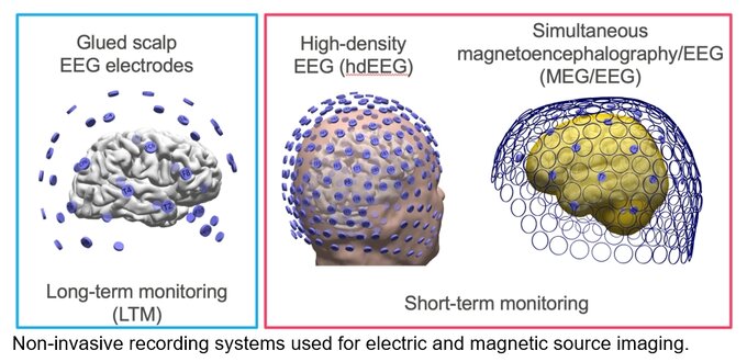
Source imaging aims at reconstructing the electromagnetic brain activity. Electrical fields or magnetic fields are picked up with electrodes at the scalp or by ultra-sensitive coils of magnetocephalography (MEG) systems, respectively. Source imaging became a valuable tool of presurgical diagnostics. It considers the conductivity profile of the patient’s individual anatomy, may involve recording systems with a high spatial sensor density and inverse solution which model focal and distributed generators of epileptic activity.
High-density scalp recordings with up to 256 channels prior to the intracranial EEG diagnostics are the basis for highly accurate source reconstruction of interictal epileptiform discharges with established and novel inverse modelling algorithms e.g. methods that are able to reconstruct the spatial extent of epileptic activity.
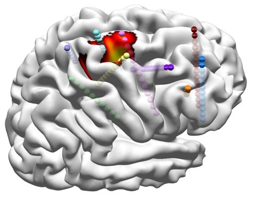
Acquisition of brain activity with intracranial electrodes offers the unique chance for the validation of source imaging results. It is further extended by simultaneous acquisition of scalp EEG. A one by one analysis of scalp and intracranial EEG signals in brain regions that contain generators of epileptic activity opens an optimal environment for mutual validation studies.
Current lines of research:
Semi-automated source imaging
Within the European Reference Network EpiCARE we further participate in the PROMAESIS trial (PI: Sándor Beniczky, Aarhus University, Denmark). The trial prospectively evaluates automatic electric source imaging algorithms of interictal and ictal epileptic activity in presurgical epilepsy diagnostics.
Heers M, Böttcher S, Kalina A, Katletz S, Altenmüller DM, Baroumand AG, Strobbe G, van Mierlo P, von Oertzen TJ, Marusic P, Schulze-Bonhage A, Beniczky S, Dümpelmann M. Detection of interictal epileptiform discharges in an extended scalp EEG array and high-density EEG - A prospective multicenter study. Epilepsia. 2022 Jul;63(7):1619-1629.
Electric source imaging of semi-automatically detected interictal epileptiform discharges using a distributed inverse method projected on the cortical surface for presurgical epilepsy diagnostics and subsequent intracerebral EEG recordings with Stereo-EEG electrodes.
Network dynamics of the electro-magnetic epileptic focus in patients with focal cortical dysplasia
In collaboration with the MEG Center, University Hospital Tübingen we evaluate network dynamics of focal cortical dysplasia in simultaneous MEG/EEG recordings during sleep. This project is funded by the DFG.
Source imaging based on invasive subdural EEG recordings (ECoG) from the brain surface
Ramantani G, Cosandier-Rimélé D, Schulze-Bonhage A, Maillard L, Zentner J, Dümpelmann M. Source reconstruction based on subdural EEG recordings adds to the presurgical evaluation in refractory frontal lobe epilepsy. Clinical Neurophysiology 124 (2013): 481–491.
Dümpelmann M., Ball T and Schulze-Bonhage, A, sLORETA allows reliable distributed source reconstruction based on subdural strip and grid recordings. Human Brain Mapping. 2012; 33:1172–1188.
Dümpelmann M, Fell J, Wellmer J, Urbach H, Elger CE. 3D source localization derived from subdural strip and grid electrodes: A simulation study. Clin Neurophysiol. 2009, 120(6):1061-9.
Multimodal simulation, imaging and inverse modelling
Cosandier-Rimélé D, Ramantani G, Zentner J, Schulze-Bonhage A, Dümpelmann M. A realistic multimodal modeling approach for the evaluation of distributed source analysis: application to sLORETA. Journal of Neural Engineering, (2017) Oct;14(5):056008.
Heers M, Hedrich T, An D, Dubeau F, Gotman J, Grova C, Kobayashi E. Spatial correlation of hemodynamic changes related to interictal epileptic discharges with electric and magnetic source imaging. Hum Brain Mapp. 2014 Sep;35(9):4396-414
Reconstructing the spatial extent of epileptic activity
Heers M, Chowdhury RA, Hedrich T, Dubeau F, Hall JA, Lina JM, Grova C, Kobayashi E. Localization Accuracy of Distributed Inverse Solutions for Electric and Magnetic Source Imaging of Interictal Epileptic Discharges in Patients with Focal Epilepsy. Brain Topogr. 2016 Jan;29(1):162-81.
Contact: PD Dr.-Ing. Matthias Dümpelmann, PD Dr. med. Marcel Heers
To learn more about …
... Source imaging in epilepsy:
https://neuroimage.usc.edu/brainstorm/Tutorials/Epilepsy
… Localization of intracranial EEG electrodes for validation of source imaging:
Abteilung Prächirurgische Epilepsiediagnostik
Ärztlicher Leiter:
Prof. Dr. Schulze-Bonhage
Breisacher Str. 64
D-79106 Freiburg
Telefon: 0761 270 53660
Telefax: 0761 270 50030
E-Mail: epilepsiezentrum@uniklinik-freiburg.de


