Microscopy Facility SCI-MED
SCI-MED (Super-Resolution Confocal/Multiphoton Imaging for Multiparametric Experimental Designs) is a new microscopy facility at the IEKM, which was established with the dedicated goal to facilitate experimental cardiovascular research in Freiburg with advanced microscopy methods and systems.
Two state-of-the-art fluorescence microscopes (a confocal and a multiphoton-/confocal) and a high-end image analysis workstation have been brought into service with financial support from the DFG (91b grant*) and the state of Baden-Württemberg*. The automated slide scanner (brightfield/fluorescence) was funded by IEKM.
An inverted confocal microscope allows recording series of optical sections and 3D imaging on various types of samples, ranging from individual cells to tissue sections, biopsies and intact organ fragments. A white light laser in combination with ultrasensitive spectral detection allows one to define fluorescence excitation lines and detection windows with nanometer-precision over a broad range of wavelengths. This supports simultaneous identification of multiple reporter molecules. The second microscope (Fig. 1B) is an upright multiphoton-/confocal microscope, used mainly for characterisation of tissue and tissue sections. This microscope allows one to detect fluorescence signals also from deeper within intact tissues, without mechanical cutting.
Both systems are equipped with a super-resolution module that employs deconvolution-based algorithms for surpassing the classic diffraction-limited resolution (x/y resolution down to 120 nm).
A special feature of the new SCI-MED systems is the possibility to also measure fluorescence lifetimes. This provides an additional data dimension, independent of colour and intensity. It enables one, for example, to reject unspecific background fluorescence, or to visualise minimal differences in the local nano-environment of a fluorescent reporter which, for instance, can be used to discriminate between healthy and diseased/injured cells.
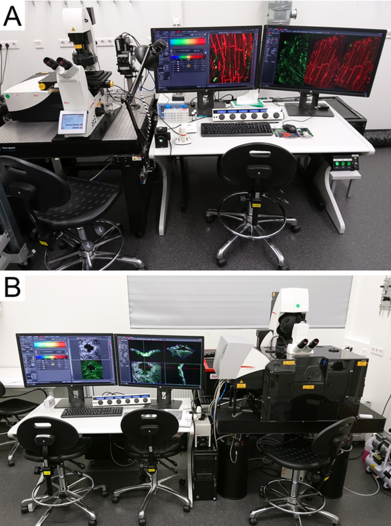
Fig. 1 The SCI-MED microsopes. A) Inverted confocal microscope: Leica TCS SP8 X. B) Upright multiphoton-/confocal microscope: Leica TCS SP8 DIVE
Detailed description of the systems
A) Inverted confocal laser scanning microscope: Leica TCS SP8 X, white light laser 470-670 nm, 405 nm laser, Argon laser (458 nm, 476 nm, 488 nm, 496 nm, 514 nm), spectral detectors (2 PMTs, 2 ultrasensitive hybrid detectors), FALCON (FLIM module), LIGHTNING (deconvolution-based super-resolution module), LAS X Navigator
B) Upright multiphoton-/confocal microscope: Leica TCS SP8 DIVE , multiphoton laser (2 lines, tunable 680-1300 nm, fixed 1045 nm), confocal (3 laser lines, 488 nm, 552 nm, 638 nm), 2 internal spectral PMT detectors, 4 non-descanned spectral detectors (2 PMTs, 2 ultrasensitive hybrid detectors), Second Harmonics Generation (SHG), FALCON (FLIM module), LIGHTNING (deconvolution-based super-resolution module), LAS X Navigator
C) Offline image analysis: HP workstation, Leica LAS-X offline analysis, ImageJ
D) Zeiss Axio Scan.Z1 (automated slide scanner), brightfield mode for histology, fluorescence mode (up to 6 channels), magazine for 100 slides
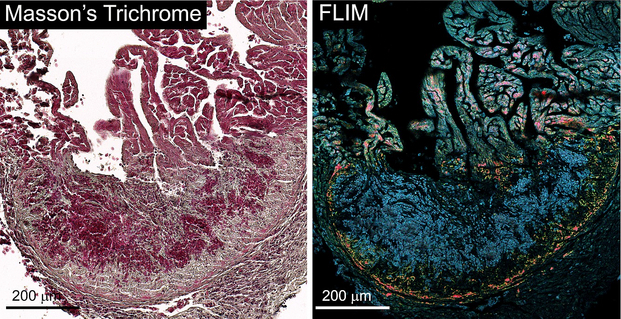
Fig. 2 Tissue section from a fish heart whose apical tissue has regenerated after cryo-injury. The histological staining (left) labels individual cells with relatively uniform colours and intensities. This makes it difficult to distinguish healthy and regenerated tissue. In contrast, fluorescence lifetime imaging of the same preparation (right) reveals significant differences in fluochrome inactivation between healthy and regenerated myocardium. Sample provided by Jennifer Esser und Martin Moser (Cardiology and Angiology I, Freiburg), microscopy was performed by Eva Rog-Zielinska (IEKM) with support from Irmtraud Steinmetz (Leica).
A short description of SCI-MED was published in the October 2018 issue of “UHZ Aktuell” (in German). The SCI-MED conditions of use are available here.
*The purchase of the microscopes was supported in equal parts by a joint major research instrumentation grant from the German Research Foundation to Peter Kohl (IEKM), Franziska Schneider-Warme/ Callum Zgierski-Johnston (IEKM), Eva Rog-Zielinska/ Remi Peyronnet (IEKM), Martin Moser/ Jennifer Esser (Cardiology and Angiology I), Ingo Hilgendorf (Cardiology and Angiology I) and Lutz Hein (Experimental and Clinical Pharmacology and Toxicology, all Freiburg University) and an infrastructure support grant from the Ministry for Science, Research and Arts in Baden-Württemberg to Peter Kohl.
Facility Manager

E-Mail: josef.madl@uniklinik-freiburg.de
Phone: +49 761 270 63953