Skull base surgery
What does a lesion in the region of the skull base mean?
The skull base includes the inferior part of the skull with its transitions to the cervical spine, the nasopharynx and facial bones. A lesion located in this region often effects the low-lying parts of the brain (eg brain stem) and the 12 cranial nerves, so that often impairment of those neural structures leads to the discovery of it.
Such a lesion can be caused by tumor (eg Sphenoid wing meningioma, acoustic neurinoma), by injury (eg fractures of the skull in the frontal skull base), by instability of the craniocervical joints or inflammatory diseases.
All images by courtesy of Prof. S. Rosahl, Erfurt.

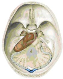
Figure 1: Meningioma in the posterior cranial base (brown). The cerebellum and brain stem can be displaced by the tumor. Tumor involving the petrous bone also grows from the posterior into the middle cranial fossa (so-called "petroclival meningiomas").
If cranial nerves are affected by a lesion, following symptoms can occur:
- Olfactory and taste disturbance (olfactory nerve)
- Visual impairment (optic nerve)
- Double vision (oculomotor nerves)
- Facial pain or numbness in parts of the face (trigeminal nerve)
- Paralysis of parts of the facial muscles (facial nerve)
- Hearing loss, tinnitus (acoustic nerve)
- Dizziness and unsteadiness (vestibular nerve)
- Swallowing disorders, hoarseness (caudal cranial nerves)
- Weakness of facial, head and shoulder muscles

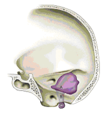
Figure 2: Tumors in the region of the caudal cranial nerves may extend through bony canals of the skull base and the foramen magnum into the region of the craniocervical junction.
Other symptoms, such as unsteadiness, paralysis and numbness throughout the body can be triggered by pressure on the brain stem (see Figure 1). Through blockage of the cerebrospinal fluid (CSF) gait disturbances, memory problems, headaches and urinary bladder dysfunction may arise.
If the anterior or the temporal lobes of the brain are compressed by a skull base lesion, personality changes and seizures may occur. Hormonal disorders (pressure on the pituitary gland) are sometimes signs of a lesion in the skull base.
Which examination method can reveal a disease in the region of the skull base?
Most lesions can be detected using magnetic resonance imaging (MRI) and der computed tomography (CT).
Often functional examinations of the cranial nerves such as hearing, eyesight and balance test are used. Electrophysiological studies (eg BAEP = brainstem auditory evoked potentials) complement these tests.
For vascular tumors sometimes vascular imaging (conventional angiography) is necessary.
What are the treatment options?
Without treatment permanent loss of function up to damage of the vital centers in the brain stem may occur.
For the successful treatment of diseases in the skull base a good collaboration of several experts in interdisciplinary centers is usually the best way. In our interdisciplinary team neurosurgeons, oromaxillofacial surgeons, ENT specialists, ophthalmologists, neuro-radiologists and radiation therapists are working together. For difficult cases the best treatment regime is being discussed in a skull base conference.
Injuries to the skull base must be cared for often very quickly. For slow-growing tumors, however, there often is no direct time pressure. Sometimes decongestant or growth-inhibiting drugs are used. Highly vascularized processes are suitable for treatment with catheters (embolisation). Radiation therapy and radiosurgery in some tumors can be very successful - often used in addition to surgery.
An operation is appropriate if other treatments are less effective, would cause severe side effects or the diagnosis is in question. The aim of the operation is the removal of the cause of the existing disease and to prevent or delay the appearance of new symptoms.
The choice of surgical method depends on the nature and location of the disease process. The surgical approach should be as minimal as possible. Particularly microsurgical and endoscopic procedures are used, sometimes in combination. Depending on the location of the disease, experts of various surgical disciplines are working hand in hand.
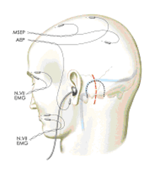
Intraoperative navigation and neurophysiologic monitoring (see Figure 3) can support the exact surgical planning and the surgery itself. Under the operation microscope, the surgeon can visualize the lesion.

Figure 3: Electrophysiological monitoring of the function of cranial nerves during surgery under general anesthesia.
Using special micro-instruments, ultrasonic aspirator, coagulation, laser or aspiration cannulae, tumors can be removed. Vascular malformations are closed with titanium clips or wrapped with various materials.
Neurosurgical methods of treatment of Graves’ disease (Morbus Basedow)
Due to this originally thyroid-associated disease, fat tissue increases within the eye-socket (orbit) caused by autoantibodies. This leads to an increased inner eye pressure which is sometimes painful, and to bulging eyes (so-called exophthalmos). Furthermore, visual impairment, diplopic images, redness, and increased sensitivity of one or both eyes may occur. In these cases, surgery – the so-called orbital decompression – may reduce eye pressure. Hereby, the orbital bone margins are partially removed through a small incision of the temple in order to provide more space for the fat tissue. This leads to a remarkable reduction of the inner eye pressure, and thus to a regression of the exophthalmos, so that the vision can recover. Diplopic images are very seldom with this method of surgery.
Consultation hours for scull base surgery
For this area we offer special consultation hours for skull base surgery.
Keywords
Trigeminal neuralgia, acoustic neurinoma, (interdisciplinary) scull base conference, scull base center, thyroid-associated (endocrine) orbitopathy, orbital decompression.

Prof. Dr. Jürgen Grauvogel, MBA
Geschäftsführender Oberarzt
Telefon 0761 270 50010
juergen.grauvogel@uniklinik-freiburg.de
Publikationen: PubMed-Liste

Dr. Christian Scheiwe, MD
Senior neurosurgeon

Dr. Christine Steiert, MD
Attending neurosurgeon
Consultation hours for skull base surgery
