Intravital Imaging Facility Freiburg
Welcome to the Intravital Imaging Facility Freiburg
The research group Eisenhardt, Clinic for Plastic and Handsurgery and the Core Facility Lighthouse offer state of the art in vivo imaging.
Recently, we established a for Freiburg one-of-its-kind possibility to perfom in vivo multiphoton microscopy. In vivo investigation of cell-cell interaction and subcellular level in small animal models (e.g. rodents) in real time is now possible.
We provide a Zeiss LSM 980 confocal-microscope with Airyscan 2. The microscope is fitted with a LED light source for single photon excitation by fluorescence microscopy Colibri 7 system (UV-line 385/30 nm, B-line 469/38 nm, and G-line 555/30 nm), and a Chameleon Discovery wide-range tunable laser system for multiphoton excitation.
Principipal investigator Prof. Steffen Eisenhardt and his team work on different DFG-funded projects. Ischemia/reperfusion injury and rejection of vascularized allograft transplants (VCA).
For further informations please find the following website.

Zeiss LSM 980 with Airyscan 2, next generation confocal microscopy equipped with Coherent Discovery tunable laser system.
Intravital multiphoton microscopy
Intravital imaging by multiphoton microscopy (MPM) is a powerful tool in the evaluation of the molecular biology in cell-cell interaction under physiological and pathophysiological conditions. Two-photon excited (TPE) fluorescence provides for major advantages over single-photon excitation microscopy, e.g. by epifluorescence wide field microscopes or confocal microscopes: (1) Due to near-infrared excitation wavelength in TPE, tissue damage by phototoxicity is reduces, with energy per photon Ephoton= h x f. (2) Secondly, longer wavelength results in less scattering and therefore greater tissue penetration with up to millimeters range. (3) Focal plane is created by “confocality” of the laser beam not by a detector pinhole. Hence, the signal-to-noise ratio (SNR) is improved and phototoxicity is reduced. These properties make MPM and TPE a most useful tool for intravital imaging of molecular biological processes.
The Coherent Chameleon Discovery system features a wide tuning range (660 to 1320 nm), dual output laser for multiphoton excitation. Chameleon specifications with >3.6 W at 800 nm and 3.5 W at 1040 nm.
"Chameleon Discovery is a high power, one-box, wide tuning range, dual output laser for Multiphoton Excitation. Literally two lasers in one, it truly redefines the boundaries of what is possible for deep, in-vivo imaging of all popular probes along with developing red shifted calcium indicators and opsins for optogenetics. The all-new model of the Chameleon Discovery with Total Power Control adds built-in, fast power modulation to both outputs, giving full control of beam parameters into a 2P microscope and streamlining the setup process so users can concentrate on acquiring quality data, rather than optimizing optical delivery systems." - Coherent
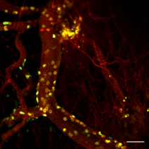
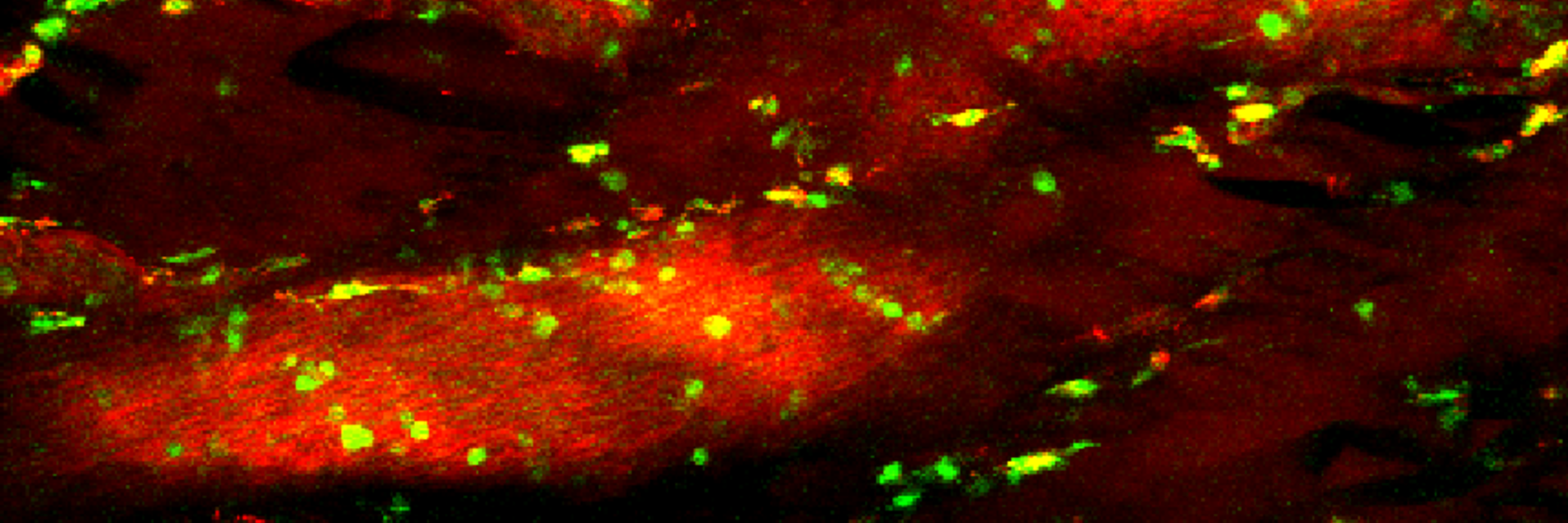
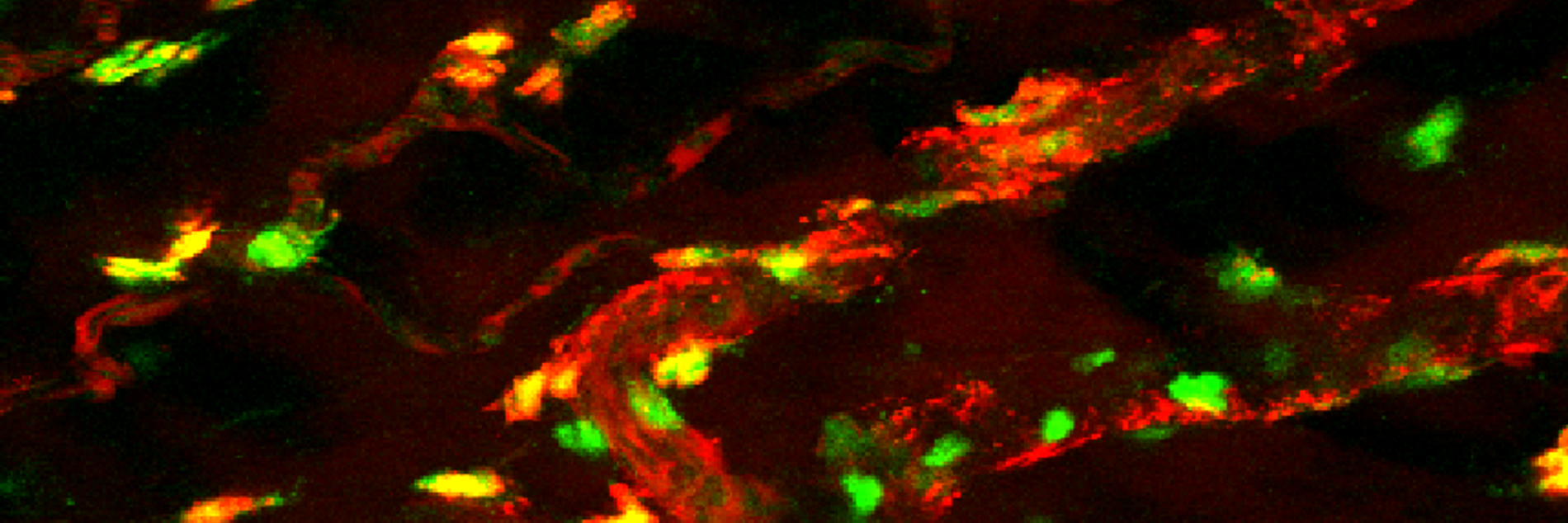
Activated CD43 (yellow) and CD68 (green) cells in the microcirulation of a transplanted hind limb in rat.
A competent partner in in vivo imaging...
Our MPM experts advice you and assist in the design and implementation of an MPM in vivo model.
With over seven years of experience in in vivo imaging and models, we provide our knowledge in this interdisciplinary imaging facility.
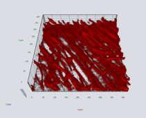
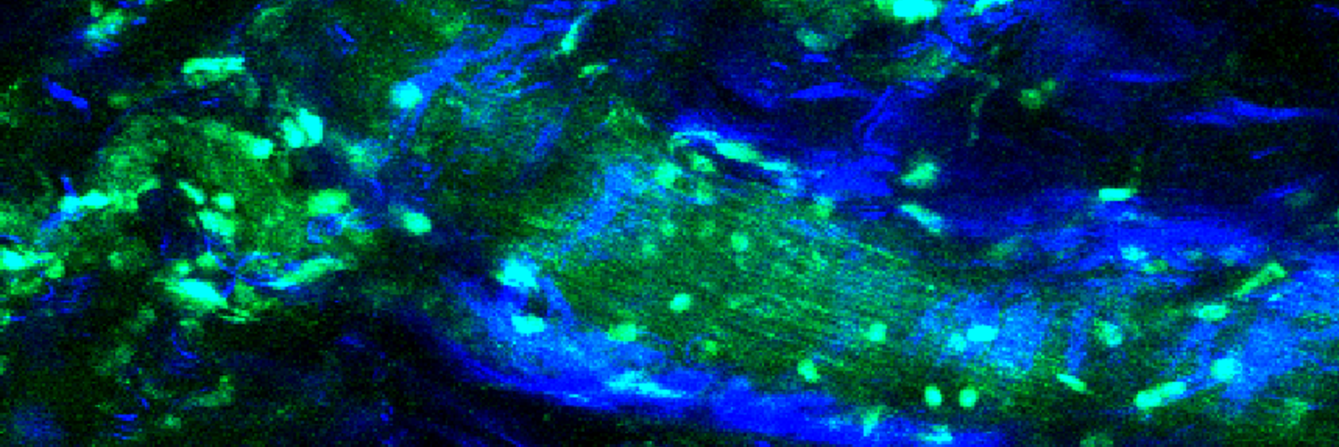
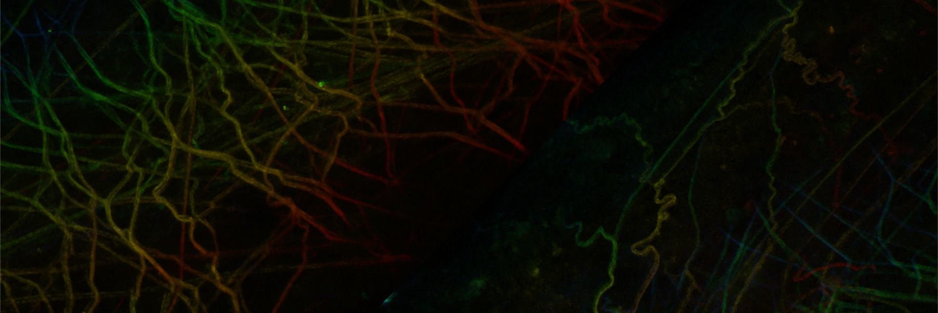
Capillary system of the tibialis anterior muscle in mouse as a 3D reconstruction.
...and in post processing
We further provide professional assistance in the post processing to help you find the best possible solution for your experimental problems.
The Freiburg Intravital Imaging Facility is funded by the German Research Foundation.
Booking system and further information
| Central booking system | link | Central booking system for reservation of the MP-microscope. Introduction needed |
| Spectra Database | link | Hosted at the University of Arizona. Helpful tool for selection of single- and multi-photon dyes |
| Life Imaging Center (LIC) | link | Microscopy and Image Analysis Platform (miap) in Freiburg. Highly competitive joint network founded 2016 with international core facilities and life science sites in Freiburg (Germany), Basel (Switzerland), Mulhouse and Strasbourg (France). |
| Custom hindlimb restrainer | link | Fixation device for artefact-free intravital imaging of the hindlimb in rodents. |
| Common fluorophores in TPE | link | Handy list of common multi photon dyes put together by Zeiss |
| Excellent instructions for measurement of velocity and diameter of multiple vessels | link | Two-Photon Imaging of Blood Flow in the Rat Cortex, Driscoll JD et al., Cold Spring Harb Protoc, 2013. |
Hugstetter Straße 55
79106 Freiburg
Telefon: 0761 270-27790
Montag-Freitag 10:00 - 13:00 Uhr