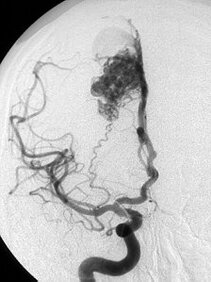
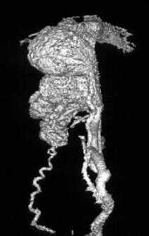
Arteriovenous malformation (AVM)
The arteries transport oxygenated blood to the organs, in this case to the brain. The venes dissipate the used blood after the oxygen exchange took place through small vasculature, the so-called capillary bed.
In case of an arteriovenous malformation, also known as angioma, this oxygen exchange is missing, the blood is running through a short-circuit connection (so-called nidus). In the neuro-imaging, this is shown as a vascular conglomerate.
In most cases, an AVM becomes symptomatic through an intracerebral bleeding. The bleeding risk is about 2-4% yearly. Further possible symptoms are seizures, headaches and neurological deficits.
Mainly due to the high bleeding risk, the AVM has to be eliminated. To achieve this aim, nowadays, an interdisciplinary center with highly specialised equipment is absolutely required in order to be able to offer an individually optimised treatment to patients.
The different methods of treatment are micro-surgical resection (neurosurgery), endovascular embolisation through catheter (neuroradiology) and radio surgery (stereotactic neurosurgery, radiation therapy) which are generally accepted. They can be employed solitary or in combination, depending on the angioms’ structural and anatomic conditions on one hand, and on the symptoms on the other hand.
Microsurgical resection
Open surgery is performed under general anesthesia and aims at removing the whole angioma at once. Hereby, an immediate elimination of the bleeding risk can be achieved. However, this method of treatment might become a high-risk surgery if the AVM is located in deeper and eloquent brain areas, if it is quite big and if it shows a complex architecture and a deeply located venous drainage.
Endovascular treatment
This treatment is performed under general anesthesia as well. Under fluoroscopy, a catheter is introduced into the femoral artery until the arteriovenous malformation is reached which is then closed up with different materials. This treatment does not require an opening of the skull.
However, in many cases the angioma cannot not be eliminated completely with only one session, therefore, either several sessions, open surgery or radio surgery are additionally necessary in order to definitely eliminate the angioma.
Radio surgery
For the treatment of arteriovenous malformation, this is basically the method with the least stress for the patients. It is a highly dosed one-time radiation which is exactly located on the angioma and can optionally be performed without anaesthetic.
This method, however, is limited to smaller angiomas, and a definitive elimination of the bleeding risk is not to be expected before 2 years after treatment.

Dr. Christian Scheiwe
Senior neurosurgeon
Consultation hours for vascular deseases

Dr. Mukesch Shah
Senior neurosurgeon


