DKTK Program
Radiation Oncology & Imaging (ROI)
Activities Partner Site Freiburg
Modern radiotherapy provides researchers with dose-space/volume-time resoluted data of individual patients which, if integrated with detailed clinical baseline and outcome data, is a powerful research tool to evaluate dose-space/volume-time responses in tumors and normal tissues. Predictive models derived from these evaluations can be translated into innovative studies on personalized radiotherapy, e.g. for biologically based inhomogeneous dose distributions which may reduce treatment sequels or increase the chance to locally control tumor.
Dpt. of Radiation Oncology
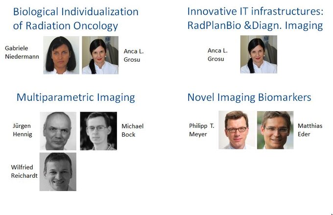
Prof. Dr. med. Anca L. Grosu
Partner Sites: All other 8 sites.
The main objective of the project is the evaluation of functional imaging for individualized radiotherapy. There are initiated and running preclinical and clinical studies to validate PET and MR-based bio-imaging methods for patient stratification in radiation dose escalation and dose-painting, target volume definition and monitoring during and after the radio-oncological treatment (see also associated studies: GLIAA, Hipporad, PET-Plan, STRIPE, LungTech, α-RT).
Freiburg furthermore coordinates in the frame of the RadPlanbio platform-project the evaluation of functional MR and amino acid PET for radiotherapy planning, monitoring and treatment evaluation in patients with glioblastoma. Another question which has to be answered within this project is the definition of pseudo-progression / pseudo-remission of the GBM after radiochemotherapy with amino acid PET. Furthermore the focus of the scientific evaluation is on the prognostic and predictive value of metabolic and hypoxic imaging of patients with head and neck cancer with primary radiochemotherapy.
Dpt. of Radiation Oncology
Prof. Anca L. Grosu
Partner Sites: All other 8 sites.
In this project, the following milestones will be implemented:
We have generated patient-derived glioblastoma (GBM) stem cell lines and found that individual GBM stem cell lines are differentially sensitive to ionizing radiation. One major aim of our work is to identify clinically applicable radiosensitizers of cancer stem cells In vitro and in orthotopic xenograft tumor models in vivo. In addition, we are interested in the development of novel tracers for the noninvasive PET imaging of cancer stem cells. Such tracers may allow the selection and the design of individually suited, molecularly-targeted therapies and may be useful for radiotherapy treatment planning and response monitoring.
Dpt. of Radiation Oncology
Prof. Dr. med. Gabriele Niedermann
Many types of tumors are organized as cellular hierarchies founded on undifferentiated tumor stem cells, commonly called cancer stem cells (CSCs). Using patient-derived CSCs, we and others have shown that only CSCs, but not differentiated tumor cells, can initiate tumors in xenograft tumor models and that CSCs are usually highly resistant to conventional chemo- and radiotherapy. We recently reported the first clinically relevant tracer for noninvasive imaging of CSCs. This tracer allows high-sensitivity and high-resolution detection of AC133+ CSCs by positron emission tomography even in orthotopically growing patient-derived glioblastoma xenografts. Based on this work, we are developing a theranostic agent based on the AC133 monoclonal antibody, that allows both the detection of AC133+ CSCs by near-infrared fluorescence imaging and their eradication by highly specific targeted infrared photoimmunotherapy (PIT). Currently, we are evaluating CSC-specific PIT for the treatment of invasively growing, CSC-driven brain tumors. We will also evaluate whether CSC-specific PIT and radiotherapy are synergistic and to what extent PIT or combinations of PIT and local tumor irradiation synergize with recently developed, clinically relevant immunotherapeutics.
Dpt. of Nuclear Medicine
Prof. Dr. med. Philipp T. Meyer & Prof. Dr. rer. nat. Helmut Mäcke
(Partner sites: Berlin, Dresden, München, Tübingen)
1. Radiosensitizing by LSD1-Inhibition:
Prostate cancer is one of the leading causes for cancer death in men. Novel therapeutic strategies are needed in the light of a rising incidence of this disease. Targeted radionuclide therapy in combination with LSD1-Inhibition is a promising therapeutic approach to achieve a strong anti-tumor effect while minimizing possible side-effects. LSD1-Inhibition causes impairment of DNA-repair mechanisms. This leads to an increased sensitivity for DNA-damaging procedures such as irradiation by targeted radionuclide therapy. Our goal is to show this effect in-vitro in cell culture, using innovative methods such as real time cellular analysis (RTCA) for different prostate cancer cell lines, and in-vivo, using appropriate xenograft models and small animal PET-imaging.
2. GLP-receptor imaging:
Most of the tracers available for imaging of GLP-1 receptor expression in pancreatic ß-cells and in insulinoma tumors are agonists, based on exendin-4 peptide. Since GLP-1 receptor agonists induce unwanted insulin secretion downstream to receptor activation, GLP-1 receptor antagonist agents would have a clear advantage as an imaging agents. Our goal is to find GLP-1 receptor antagonist, which can be suitable for imaging of insulinomas and potentially for measuring pancreatic ß-cell mass.
3. GIP-receptor imaging:
A new family of peptide receptors, the incretin receptor family, overexpressed on many neuroendocrine tumors (NETs) is of great importance because it may enable the in vivo peptide-based receptor targeting of a category of NETs which do not express the somatostatin receptors. This project aims at developing and evaluating a new class of radioligands with the potential to be used for the in vivo targeting of GIP-receptor positive tumors. A library of GIP ligands based on the truncated peptide GIP(1-30) was develpoed with the potential to be used for the imaging of a broad spectrum of NETs (EG2 to EG8). The preclinical evaluation of 68Ga-EG4 and 68Ga-EG6 indicated feasible diagnostic imaging of GIPr-positive tumors. The good characteristics of these radiotracers support their potentiality as PET imaging probes for a broad spectrum of NETs and this allows us to further continue in the development of this family of radioligands.
4. New potent bombesin-based antagonists for cancer imaging and radionuclide therapy:
The gastrin-releasing peptide receptor (GRPr) appears to be an important molecular target for the visualization of prostate, cancer and ovarian cancers and is currently being targeted with radiolabeled bombesin derivatives. Bombesin-based agonists have been developed and evaluated in (preclinical and) clinical studies. Besides having inferior pharmacokinetics they showed serious side effects. We therefore initiated a research project aiming at developing antagonistic GRPr radioligands for clinical translation. We developed several very potent GRPr-based antagonists. They show promising in vivo pharmacokinetics and may contribute to the improvement of the diagnostic imaging of tumors overexpressing GRPr. One of them is currently being studied in a phase I study. We continue on the development of GRPr-based radioligands by investigating several chelating systems, introducing various N-terminal modulations or structural modifications on the GRPr-radioantagonists aiming at further improving the pharmacokinetics.
In addition to studies in patients with prostate cancer, we conducted first GRPr PET examinations in patients with locally advanced breast cancer on compassionate-use basis. These studies provided very promising results. In line with the literature, our preliminary data suggest that GRPr expression is strongly associated with estrogen and progesterone receptor status. In addition to these human in vivo studies, we are performing additional in vitro studies to unravel des relationship between GRPr and hormone receptor expression in breast cancer. This knowledge will help to properly interpret the imaging results and potentially may aid patient selection and outcome prediction in anti-estrogen therapies.
5. Development of peptidomimetic urea-based prostate-specific membrane antigen (PSMA) inhibitors:
Efforts to evaluate and discover diagnostic and therapeutic markers for prostate cancer continue. Recent clinical experiences suggest that the prostate-specific membrane antigen (PSMA), a transmembrane protein expressed in all types of prostatic tissue, represents a very promising diagnostic and therapeutic target. Several low molecular weight inhibitors of PSMA have been developed, radiolabeled with several radionuclides and investigated as imaging agents in murine models of prostate cancer. Amongst them, 68Ga labeled Glu-NH-CO-NH-Lys(Ahx)-HBED-CC is currently under clinical assessment showing impressive results. Within the frame of this project we aim at developing improved PSMA-based inhibitors as PET tracers for the non-invasive imaging of prostate cancer. The urea-based peptidic analog NODAGA-Phe-Phe-D-Lys(suberoyl-Lys-urea-Glu) (VG66) is currently under investigation. VG66 is labeled with 68Ga and 64Cu. The in vitro evaluation of the labeled compounds which includes internalization and saturation binding studies showed high affinity of 68Ga-VG66 and 64Cu-VG66 (with the PSMA positive cell line LNCaP). PET imaging and pharmacokinetic studies show very promising results outperforming literature data.
6. Hybrid ligands targeting prostate cancer for imaging (PET), targeted radionuclide therapy and intraoperative localization:
It has been shown that on the plasma membrane of prostate tumor cells there are two highly overexpressed molecular targets: the prostate specific membrane antigen (PSMA) and the gastrin-releasing peptide receptor (GRPr), a G-protein coupled receptor (GPCr) of the bombesin receptor family. There are also indications that the GRPr has lower expression at high Gleason score whereas PSMA appears to remain highly expressed in CRPC. The simultaneous coexistence of PSMA with GRPr on prostate tumor cells might argue for multiple receptor targeting using hybrid ligands.
Dpt. of Radiology / Medical Physics
Prof. Dr. Dr. h.c. Jürgen Hennig & Prof. Dr. Michael Bock
Dr. Wilfried Reichardt
(Partner sites: Dresden, Essen/Düsseldorf, Heidelberg, München, Tübingen)
The project is developing novel MR imaging methods for the early diagnosis, differentiation and follow-up during treatment of the glioblastoma. In particular, in a patient study at 3 Tesla the use of time-of-flight MR angiography is evaluated for the characterization of the arterial vascular supply of glioblastomas. To unravel the complex underlying contrast mechanisms of the time-of-flight signal in the irregular neo-angiogenic vascular structures in tumors, a small animal study is currently initiated in a mouse model. In addition, diffusion and oxygen MRI techniques are developed to assess glioblastoma heterogeneity and oxygen consumption, and image post-processing methods are implemented to quantitatively describe changes of tumor vasculature under anti-angiogenic treatment.
The MR methods developed in this methodological research are transferred also to other tumor entities. Currently, in Freiburg several clinical studies are starting or are planned, in which diffusion MRI, oxygen-sensitive imaging and perfusion quantification are combined with conventional MR imaging of different tumor entities such as prostate carcinomas or head-and-neck tumors.

