Experimental Radiology
Advanced Imaging ConceptsMRI has several limitations such as the limited signal-to-noise ratio or the reduced patient access in the large magnets. To overcome these limitations we create advanced imaging concepts that go beyond the current MRI technology: we develop a purely optical RF coil design (“light coils”), we use MRI to measure radiation doses (gel dosimetry), and we extend the clinical applications to new fields such as dental MRI. In addition, we use artificial intelligence to improve the lesion detection based in oncology.
In this project, deep neural networks are developed to process and analyze extensive patient data to enhance the automatic segmentation of head, neck, and prostate tumors. By integrating AI research with MRI cancer imaging, imaging protocols are improved and patient treatment is optimized. Explainable AI techniques are employed to ensure transparency and interpretability in the models, and data fusion techniques are used to integrate diverse data sources for more accurate analysis. This work connects AI with the optimization of MRI techniques and comprehensive data processing.
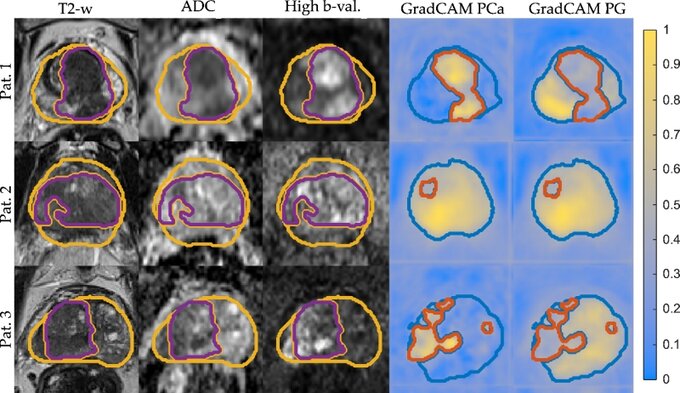
Figure 1: Segmentation of PG and PCa for test patient 1–3 with the corresponding input mpMRI sequences and ground truth labels PG (yellow) & PCa (purple). The corresponding Grad-CAM maps are overlaid with the network predicted segmentation for PG (blue) & PCa (orange). Figure from ref. 2.

Figure 2: Segmentations of GTV overlaid on the input image sequences for patients from the test set. Ground truth segmentations PCa-Histo (purple), PCa-Rad (blue) and the predicted segmentation PCa-CNN (orange). Figure from ref. 1.
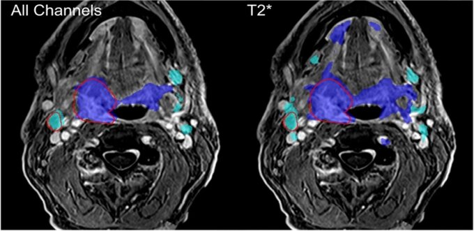
Figure 3: Segmentation result for the reference CNN and the LOO-CNN without T2* input. A distinct over segmentation is observed in both cases, which is much less pronounced in the reference CNN. In this image the reference CNN has a DSC of 72% / 45% for GTV-T / GTV. Figure taken from ref. 3.
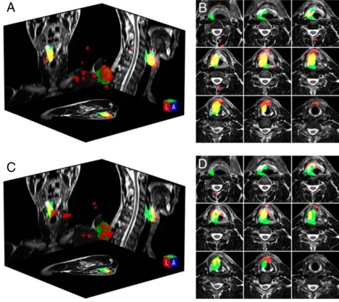
Figure 4: 3D visualization of a segmentation with (A) and without (C) additional distortion-corrected diffusion input data. Corresponding transverse slices of the region of interest are shown (B, D). The ground truth is shown in green, and the segmentation results. Figure taken from ref. 4.
References
Gunashekar, D.D., Bielak, L., Oerther, B. et al. Comparison of data fusion strategies for automated prostate lesion detection using mpMRI correlated with whole mount histology. Radiat Oncol 19, 96 (2024).
Gunashekar, D.D., Bielak, L., Hägele, L. et al. Explainable AI for CNN-based prostate tumor segmentation in multi-parametric MRI correlated to whole mount histopathology. Radiat Oncol 17, 65 (2022).
Bielak, L., Wiedenmann, N., Berlin, A. et al. Convolutional neural networks for head and neck tumor segmentation on 7-channel multiparametric MRI: a leave-one-out analysis. Radiat Oncol 15, 181 (2020).
Bielak L., Wiedenmann N., Nicolay N.H., Lottner T., Fischer J., Bunea H., Grosu A.-L., Bock M. Automatic Tumor Segmentation With a Convolutional Neural Network in Multiparametric MRI: Influence of Distortion Correction. Tomography. 2019; 5(3):292-299.
Cooperation Partners
Department of Radiation Oncology, Freiburg University and Faculty of Medicine, Freiburg, Germany
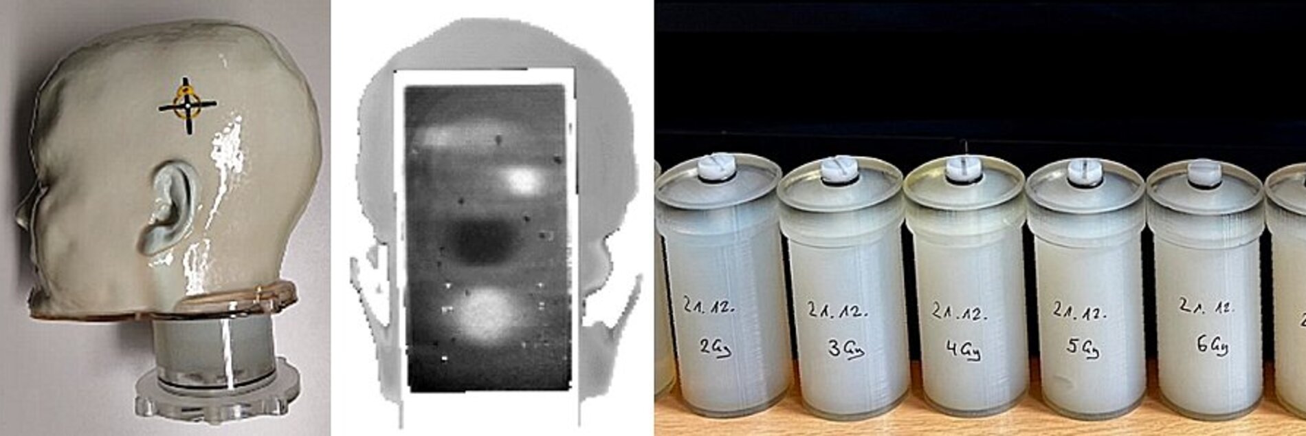
For optimal accuracy in radiation therapy, tumors are positioned in the center of the radiation system. Treatment of multiple metastases thus requires time-consuming repositioning for each metastasis. In this project we use faster off-center irradiation of multiple metastases and investigate how experimental factors (e.g., radiation-therapy device, collimator setting) influence the accuracy of the irradiation. We use radio-sensitive polymer gels that experience an increase of R2 relaxation rates under irradiation. From these changes, we can calculate the locally delivered dose and compare to the expected radiation plan. The measurements are performed as a multicenter study in the DACH region.

Cooperation Partners
Department of Radiation Oncology, Freiburg University and Faculty of Medicine, Freiburg, Germany
Grant support
Funded by the German Cancer Aid (Deutsche Krebshilfe) under grant number 701143317: “Untersuchung der Anforderungen an Sicherheit, Genauigkeit und Qualitätssicherung bei der Stereotaktischen Bestrahlung Multipler Hirnmetastasen mit einem Isozentrum”
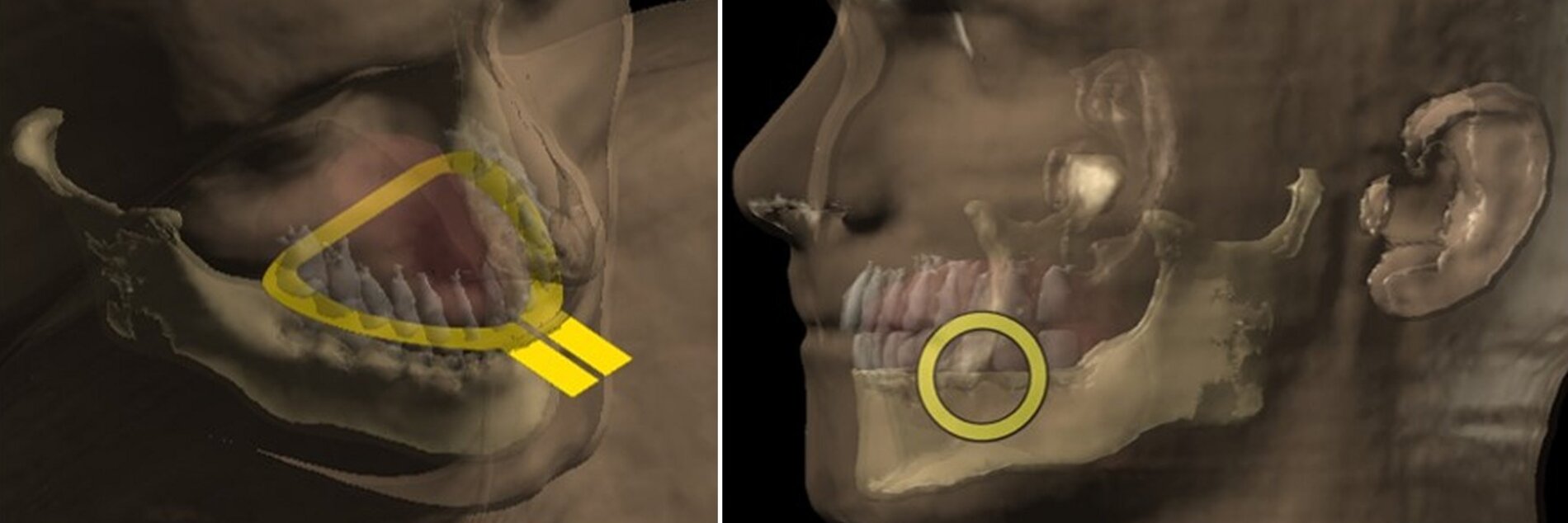
In a conventional MRI exam the excitation and acquisition events are time-interleaved: MR signal is first generated by a strong radio-frequency excitation and later the weak MRI signal is acquired. Concurrent excitation and acquisition (CEA) MRI is performed by simultaneous excitation of the spins and acquisition of the resulting signal. Concurrent excitation and acquisition (CEA) can overcome these limitations, but CEA is extremely challenging due to the huge difference between transmit and receive signal levels. Therefore, we developed novel techniques to decouple transmit and receive parts of the MRI system, and adapted full-duplex telecommunication techniques to MRI to implement CEA in clinical MRI systems.
We have developed a novel software tool for optimization of the CEA contrast. With CEA the signal of ultrashort-T2* species has become detectable. We demonstrated nearly 100 % MR signal acquisition efficiency by CEA. In addition, CEA can be eliminated with virtually no acoustic noise, thus increasing patient comfort. Finally we have used extremely low peak RF powers and reduced the RF-power-deposition-related risks of MRI. CEA MRI is a promising tool for various clinical applications including musculoskeletal imaging, dental imaging, connective tissue and myelinated neurons.
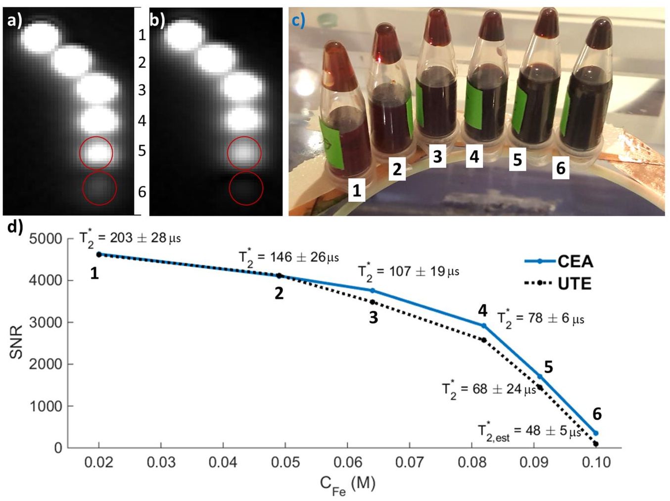
Top: Comparison of CEA and UTE in MRI of iron-oxide solutions. A coronal slice from the images acquired with (a) CEA and b) UTE. (c) A photo of the iron-oxide phantoms with different concentrations placed on the CEA coil form, which is made of glass. (d) SNR comparison for various Fe concentrations and T2* values measured using the UTE sequence. The last T2* value, i.e., for phantom 6, was estimated by assuming linear dependence of 1/T2* on CFe. From: Özen AC et al. Sci Rep 8, 10631 (2018).
References
Özen, A. C., Bock, M., & Atalar, E. (2015). Active decoupling of RF coils using a transmit array system. Magnetic Resonance Materials in Physics, Biology and Medicine, 28(6), 565–576.
Özen, A.C., Atalar, E., Korvink, J.G., Bock, M. In vivo MRI with Concurrent Excitation and Acquisition using Automated Active Analog Cancellation. Sci Rep 8, 10631 (2018).
Cooperation Partners
University of Minnesota, Center for Magnetic Resonance Research
Johannes Fischer, Agazi Tesfai
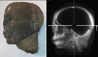
This project focuses on MR imaging of ancient human remains, which poses two main problems: The artificial dehydration necessary for the successful preservation vastly reduces signal strength and the increased coupling of the remaining hydrogen nuclei in the solid material leads to very short T2* relaxation times. For imaging of these specimens we use ultra-short echo time (UTE) and single point imaging (SPI) sequences as well as custom built transmit/receive coils. The coils are simulated and adjusted to the sample with analytic estimation of signal to noise ratio (SNR) depending on coil geometry and specimen.
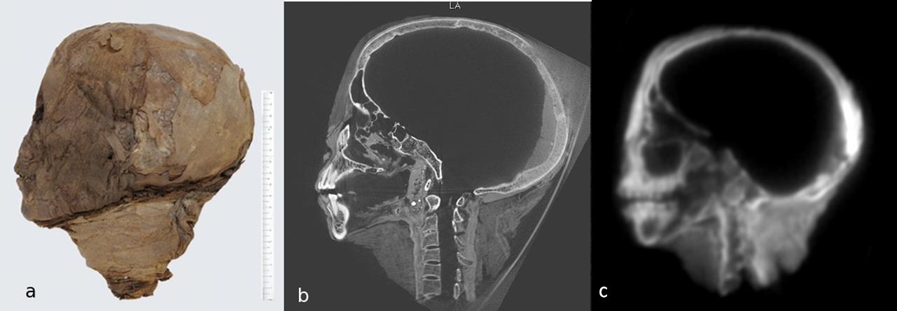
Figure 1: a) Egyptian mummy head as measurement sample, b) corresponding CT image, and c) MR image acquired with a custom built Birdcage coil and UTE sequence.
References
Tesfai, A.S., Fischer, J., Özen, A.C., Eppenberger, P., Oehrstroem, L., Rühli, F., Ludwig, U. and Bock, M., 2020. Multi-parameter Analytical Method for B1 and SNR Analysis (MAMBA): An open source RF coil design tool. Journal of Magnetic Resonance, 319, p.106825.
Özen, A.C., Ludwig, U., Öhrström, L.M., Rühli, F.J. and Bock, M. (2016), Comparison of ultrashort echo time sequences for MRI of an ancient mummified human hand. Magn. Reson. Med., 75: 701-708.
Fischer, J. Özen, A.C., Kurzhunov, D., Reisert, M., Tesfai, A., Rühli, F.J., Ludwig, U. and Bock M. Cross-Modality MR Image Reconstruction: CT-Constrained Anisotropic Diffusion to Preserve Edge Information in MRI of an Ancient Mummified Hand, Proceedings of the 25th ISMRM annual meeting, Honolulu, Hawaii.
Tesfai, A. Fischer, J., Özen, A.C., and Bock M. Comparison of different RF coil designs for short T2* samples, Proceedings of the 25th ISMRM Annual Meeting, Honolulu, Hawaii.
Tesfai, A. Fischer, J., Özen, A.C., Ludwig, U. and Bock M. Effects of Reduced Dead Times on the SNR of Tissue with Tissue with Short T2* values, Joint Annual Meeting ISMRM-ESMRMB, Paris, France.
Tesfai, A. Fischer, J., Özen, A.C., Ludwig, U. and Bock M. Multiparameter Analysis Method for B1 Acquisition (MAMBA): A tool for RF coil design and SNR estimation for short T2* samples, Proceedings of the 27th ISMRM Annual Meeting, Montréal, QC, Canada.
Cooperation Partners
Institute of Evolutionary Medicine (IEM), University of Zürich
Grant Support
Deutsche Forschungsgemeinschaft (DFG) Grants BO 3025/8-1 and LU 1187 / 6-1
Imaging techniques for MRI of lung dynamics under different patient postures (e.g., in lying and weight-bearing positions) using a rotatable 0.25 T open MR system (in coop. with ESAOTE)
MRI pulse sequences for imaging of ancient remains such as Egyptian mummies (in cooperation with the Univ. Zürich)

(a) Left hand of the Egyptian mummy
(b) Mummy hand placed in the solenoid coil developed at the Department of Experimental Radiology
References
Özen A, et al. Comparison of ultrashort echo time sequences for MRI of an ancient mummified human hand. Magn Reson Med. 2015 Mar 7. [Epub ahead of print]

Prof. Dr. Michael Bock
Director of Experimental Radiology
Tel.: +49 761 270-94140
E-Mail: michael.bock@uniklinik-freiburg.de
University Medical Center Freiburg
Dept. of Radiology · Medical Physics
Killianstr. 5a
79106 Freiburg

Oksana Chikh
Administrative Assistant
Tel.: +49 761 270-93840
E-Mail: oksana.chikh@uniklinik-freiburg.de