Experimental Radiology
Functional High Field MRITo assess tissue function, we develop novel imaging pulse sequences and hardware for MRI at very high fields (B0 ≥ 3 T). Tissue oxygenation, metabolism and inflammation are studied using non-proton MRI techniques, and the oscillation of the vocal folds is imaged using novel MRI techniques with sub-millisecond temporal resolution. For the imaging experiments both the small animal imaging systems (7 T and 9.4 T Bruker) at AMIR and a 7 T whole body MR system at the DKFZ are used.
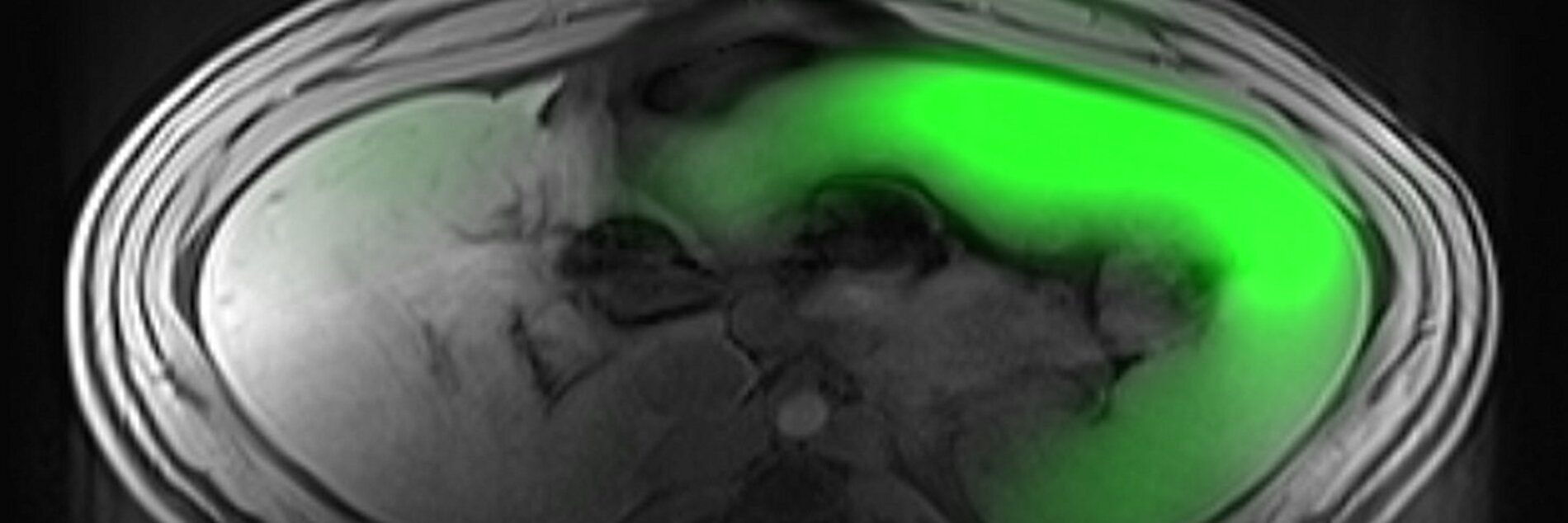
Myeloid cells are involved in inflammatory healing processes during and after myocardial infarction and are therefore of interest for novel therapeutic approaches. Since they take up injected perfluorocarbons (e.g. perfluorooctyl bromide (PFOB)), these cells can be tracked using 19F MRI. This project focuses on developing 19F MRI imaging methods to maximize the signal gained from such tracers.
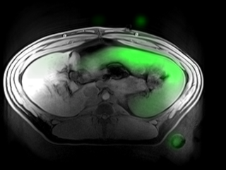
Combined 1H and 19F image of a porcine spleen after PFOB injection
Cooperation Partners
References
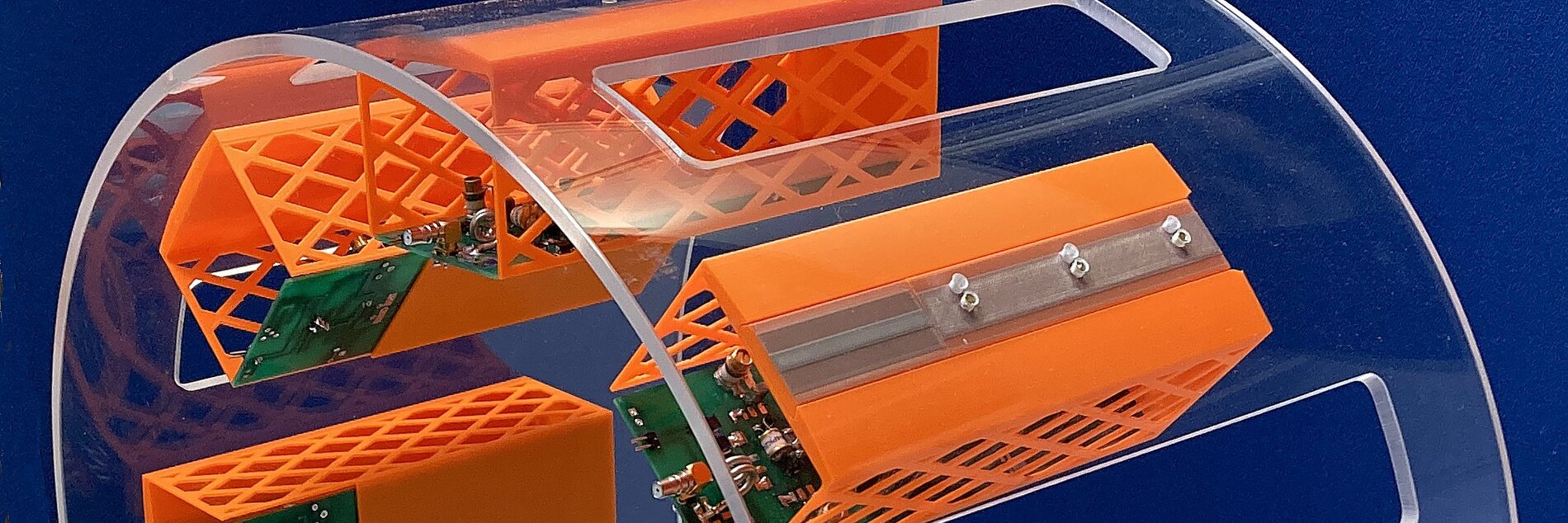
Özen AC, Spreter F, Schimpf W, et al. Scalable and modular 8-channel transmit and 8-channel flexible receive coil array for 19 F MRI of large animals. Magn Reson Med. 2023;89(3):1237-1250.
Johannes Fischer, Paula Jordan
Conventional optical laryngoscopy is limited to a top down view of the vocal fold surface. This makes it insensitive to important physiological features such as the vocal fold contact area or the vertical oscillation amplitude. To reveal this dynamic behavior below and within the vocal folds, we develop MRI techniques than can resolve this rapid motion with sub-millisecond temporal resolution, by using time-optimized MRI pulse sequences. We have successfully imaged the motion in 1D and 2D and are currently working on a fully isotropic 3D implementation.
References
Özen, A.C., Traser, L., Echternach, M., Dadakova, T., Burdumy, M., Richter, B. and Bock, M. (2016), Ensuring safety and functionality of electroglottography measurements during dynamic pulmonary MRI. Magn. Reson. Med., 76: 1629-1635.
Fischer, J, Abels, T, Özen, AC, Echternach, M, Richter, B, Bock, M. Magnetic resonance imaging of the vocal fold oscillations with sub‐millisecond temporal resolution. Magn Reson Med. 2020; 83: 403– 411.
Fischer J, Özen AC, Ilbey S, Traser L, Echternach M, Richter B, Bock M. Sub-millisecond 2D MRI of the vocal fold oscillation using single-point imaging with rapid encoding. MAGMA. 2022 Apr;35(2):301-310.
Cooperation Partners
Institute for Musicians’ Medicine, Freiburg University and Faculty of Medicine, Freiburg, Germany
Grant Support
Funded by DFG project number 525074946.
Hao Song, Jeffry Omega Prima, Ali C. Özen
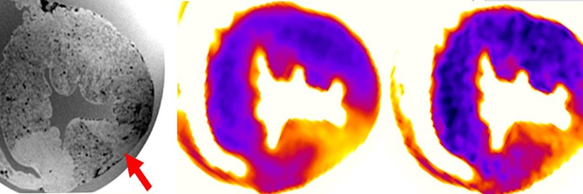
Direct 17O-MRI can be used to observe the dynamics of renal metabolism in kidney transplants. In this work 17O-MRI methods are developed in a porcine kidney in an organ transplantation setup at 3T. To obtain stable SNRs above 20 over time while maintaining a spatial resolution below 8 mm, we investigated the influence of nominal spatial resolution, bandwidth and acquisition time window of a UTE-sequence with golden-angle acquisition pattern on SNR. Signal increase of up to 25% per liter of 17O-gas was observed in a pilot experiment.
To increase SNR further, a dedicated 17O Rx array was designed that fits into a perfusion box used for functional metabolism tests of the donor kidneys. The increased filling ratio resulted in higher SNR compared to the volume and surface Tx/Rx coils. In combination with a Tx-only birdcage 17O coil for homogeneous excitation, the 4-element Rx array also enables parallel imaging.
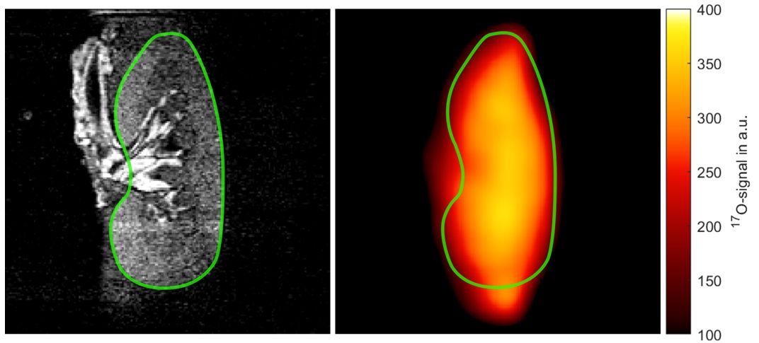
Top: Anatomical, isotropic 1H-MPRAGE image of the perfused porcine kidney (left) and the corresponding interpolated 17O-magnitude data (right), which were averaged over the entire scan time. Both images are intrinsically registered since 1H-data were acquired by the body coil while the 17O-coil was inserted.
References
Yanis Taege, Johannes Fischer, Ali Çağlar Özen, Hao Song, Christian Schuch, Rianne Schutter, Cyril Moers, Ronald JH Borra, and Michael Bock. Protocol Optimization for Functional 17O-MRI of Donor Kidneys at 3T. ISMRM 27th Annual Meeting & Exhibition 2019.
Hao Song, Ali Özen, Johannes Fischer
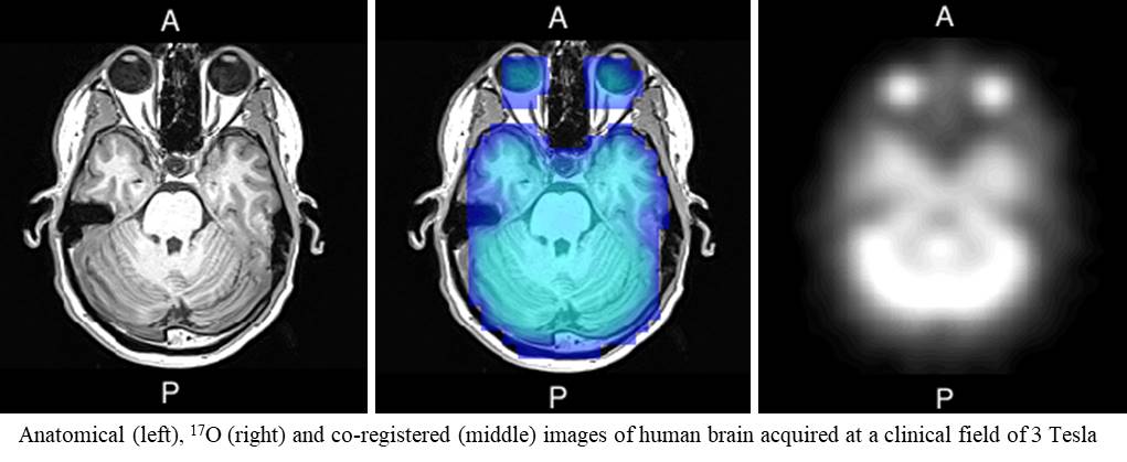
CMRO2 quantitatively represents the rate of oxygen consumption by metabolic processes in brain tissue. Dynamic 17O MR imaging is used to measure it directly and noninvasively through inhalation of isotope-enriched 17O gas (in coop. with Nukem Isotopes). The MR signal change is monitored using dedicated coil hardware, pulse sequences and post-processing methods. Currently, all measurements are performed at a clinical field strength of 3 Tesla in order to apply this technology in patients in the future.
References
Kurzhunov, D., Borowiak, R., Reisert, M., Özen, A.C., Bock, M., 2018. Direct estimation of 17O MR images (DIESIS) for quantification of oxygen metabolism in the human brain with partial volume correction. Magnetic Resonance in Medicine 80(6), 2717-2725.
Kurzhunov, D., Borowiak, R., Hass, H., Wagner, P., Krafft, A.J., Timmer, J., Bock, M., 2017. Quantification of oxygen metabolic rates in human brain with dynamic 17O MRI: profile likelihood analysis. Magnetic Resonance in Medicine 78(3), 1157-1167.
Kurzhunov, D., Borowiak, R., Reisert, M., Krafft, A.J., Özen, A.C., Bock, M., 2017. 3D CMRO2 mapping in human brain with direct 17O MRI: comparison of conventional and proton-con-strained reconstructions. NeuroImage 155, 612-624.
Borowiak, R., Reichardt, W., Kurzhunov, D., Schuch, C., Leupold, J., Krafft, A.J., Reisert, M., Lange, T., Fischer, E., Bock, M., 2017. Initial investigation of glucose metabolism in mouse brain using enriched 17O-glucose and dynamic 17O-MRS. NMR in Biomedicine 30, e3724.
Borowiak, R., Groebner, J., Haas, M., Hennig, J., Bock, M., 2014. Direct cerebral and cardiac 17O-MRI at 3 Tesla: initial results at natural abundance. Magnetic Resonance Materials in Physics, Biology and Medicine 27, 95-99.
Hoffmann SH, et al. Direct 17O MRI with partial volume correction: first experiences in a glioblastoma patient. MAGMA 2014;27(6):579-587
Cooperation Partners
Maddalena Strumia, Wilfried Reichardt
To differentiate between abnormal tumor vessels and regular brain vasculature using new quantitative measures in time-of-flight (TOF) MR angiography (MRA) data (in cooperation with the DKTK)
References
Strumia M, Reichardt W, Staszweski O, Heiland DH, Weyerbrock A, Mader I, Bock M. Glioma vessel abnormality quantification using time-of-flight MR angiography. MAGMA. 2016 Oct;29(5):765-75.
Radio-frequency coils and pulse sequence technologies for parallel signal reception and excitation (pTx) developed for both animal and human-sized MR systems (in cooperation with IMTEK and QED)

Prof. Dr. Michael Bock
Director of Experimental Radiology
Tel.: +49 761 270-94140
E-Mail: michael.bock@uniklinik-freiburg.de
University Medical Center Freiburg
Dept. of Radiology · Medical Physics
Killianstr. 5a
79106 Freiburg

Oksana Chikh
Administrative Assistant
Tel.: +49 761 270-93840
E-Mail: oksana.chikh@uniklinik-freiburg.de