Core Facility für Histopathologie und Digitale Pathologie
Die Core Facility für Histopathologie und Digitale Pathologie unterstützt als Technologieplattform durch standardisierte und qualitätsgesicherte Prozesse lokale, nationale und internationale Wissenschaftler bei der feingeweblichen Analyse von humanen Gewebeproben und davon abgeleiteten Derivaten sowie der Entwicklung innovativer gewebebasierter Methoden.
Die Core Facility für Histopathologie und Digitale Pathologie wird geleitet von Herrn PD Dr. Peter Bronsert und Herrn Professor Dr. Dr. Christoph Schell.
Bei Fragen zu Projekten, Gewebeprozessierung, Untersuchungen oder Kooperationen erreichen Sie uns über: pathologie.projektkoord@uniklinik-freiburg.de
Wir freuen uns, Ihre Projekte aus der Medizinischen Fakultät des Universitätsklinikums Freiburg, weiteren Fakultäten der Universität Freiburg und Projekte externer Kooperationspartner unterstützen zu dürfen.
Die Core Facility für Histopathologie und Digitale Pathologie unterstützt folgende Analysen:
- Makroskopische und mikroskopische Gewebebeschreibungen
- Gewebeprozessierung
- Histochemie Färbungen und Auswertungen
- Immunhistochemische Färbungen und Auswertungen
- Digitale Pathologie und digital assistierte Auswertung histochemischer und immunhistochemischer Färbungen
Leistungsangebot CFHDP
Schnitterstellung
- Leica CM 1950
- Leica RM 2255
- Leica IP C Injekt Printer Kassettendrucker
- Slice Culture / Vibratom
- HistoCore Arcardia C - Kühlplatte
Tissue MicroArray (TMA) Erstellung
Histochemie Färbung und Auswertung
(u.a. Hämatoxylin / Eosin, Periodic acid-Schiff, Elastika-van-Gieson-Färbung)
- Dako Cover Stainer
- TissueTek Prisma
- KI-unterstützte Bildanalyse mit QuPath
- Leica ST4020 kompakter linearer Färbeautomat
Immunhistochemie und Auswertung
(> 230 etablierte Färbungen)
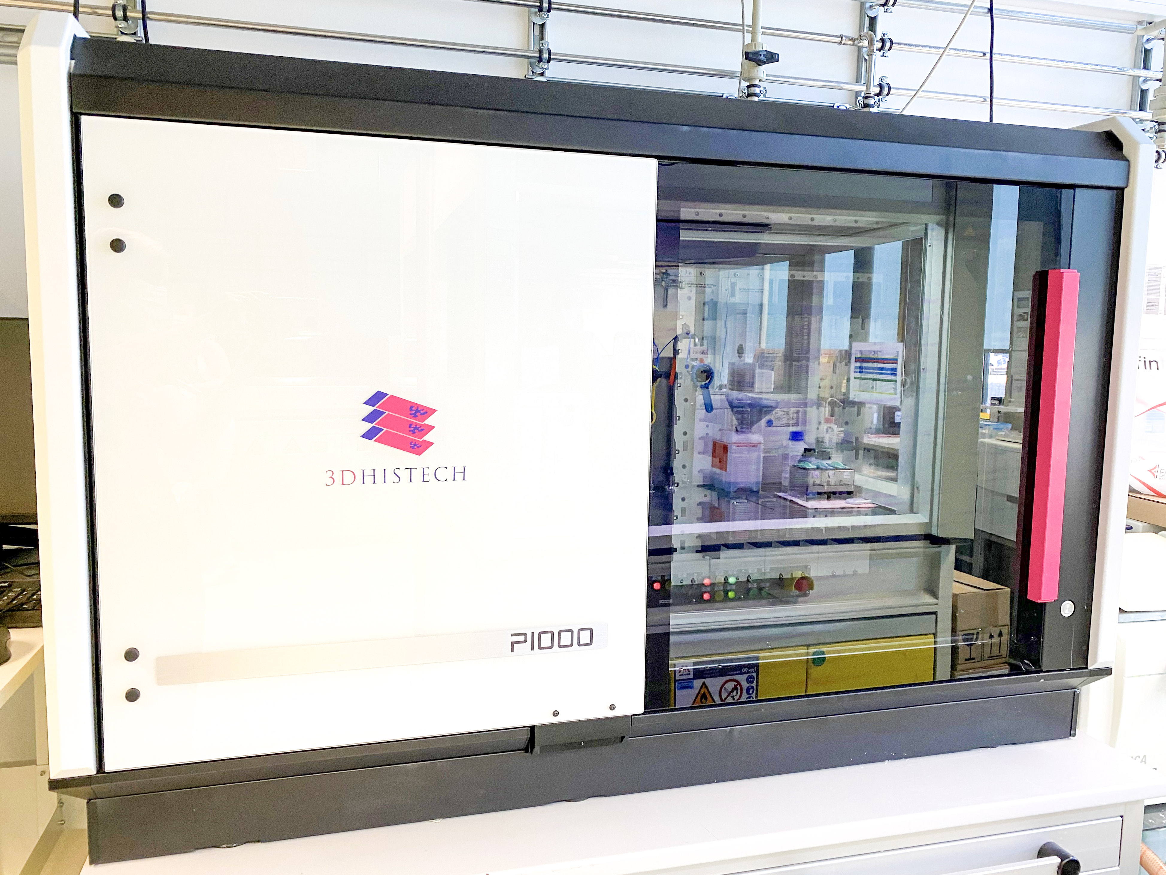
Schnittbilddigitalisierung
- 3D Histech Panoramic Scan
- Roche Ventana DP 200
- 3DHISTECH Pannoramic 1000 Flash IV
- SliceCenter - Datenbank für Ihre Gewebeschnitte
Pseudonymisierung
Im Rahmen einer- in Kooperation mit der Core Facility für Histopathologie und Digitale Pathologie- geplanten Promotion oder eines gemeinsamen Forschungsvorhabens, werden die neuen Doktorand*innen und wissenschaftliche Mitarbeiter*innen (im folgenden Mitarbeitende) umfassend in die örtlichen Strukturen und benötigten Methoden eingearbeitet. Dieses findet anhand eines festgelegten und innerhalb von roXtra (Intranet) hinterlegten Einarbeitungsplanes statt.
Gestartet wird mit dem Einarbeitungsgespräch (und ggf. der Unterzeichnung der Promotionsvereinbarung). Im Laufe der ersten Woche lernen die neuen Mitarbeitenden das Institut für Klinische Pathologie und die Core Facility für Histopathologie und Digitale Pathologie, deren Strukturen sowie Arbeitsabläufe kennen. Dieses beinhaltet u. a. auch die Einführung zu QM-relevanten Themen und zum allgemeinen Ablauf am Arbeitsplatz Labor. Anschließend werden die pathologiespezifischen Themen mit der Einführung in die IT Systeme, die Histopathologie und die individuell benötigten Labortätigkeiten gemeinsam erarbeitet.
Nach vier Wochen erfolgt das erste Zwischengespräch mit dem / der zugeteilten Tutor*in. Das Erlernte wird rekapituliert, notwendige Vertiefungen sowie aufgetretene Schwierigkeiten erörtert, Lösungen gefunden und das weitere Vorgehen abgestimmt.
Bei Bedarf werden die Mitarbeitenden in die Methoden der digitalen Pathologie eingearbeitet. Dieses beinhaltet die Handhabung unterschiedlicher Slide Scanner und KI-Bildanalysesoftware.
Nach acht Wochen findet das Abschlussgespräch, mit der Planung der nächsten Arbeitsschritte, notwendiger Anpassungen und Fortbildungen, statt. Ferner ist eine gemeinsame Evaluation der Einarbeitung vorgesehen, um diese gegebenenfalls anzupassen bzw. zu optimieren.
| Auer, Susanne | Sekretärin | ||
| Auer, Thorben | Aushilfe IT | ||
| Bronsert, Peter | PD Dr. | Leiter | |
| Eckermann, Tanja | Qualitäts- und Prozessmanagement | ||
| Kurowski, Konrad | Arzt | ||
| Schell, Christoph | Prof. Dr. Dr. | Vertreter | |
| Schmid, Marlene | Medizinisch-technische Assistentin (MTA) | ||
| Timme-Bronsert, Sylvia | Dr. | Stellvertreterin | |
| Übelin, Anja | Wissenschaftliche Hilfskraft |
|
Institution |
Arbeitsgruppe/Projektleiter*in |
|
Institut für Klinische Pathologie /UKF |
AG Schilling, Prof. Dr. Schilling |
|
AG Schell, Prof. Dr. Schell |
|
|
AG Föll, Dr. Föll |
|
|
AG Schultheis, Prof. Dr. Schultheis |
|
|
CCI |
Prof. Dr. Stephan Ehl |
|
Prof. Dr. Klaus Warnatz |
|
|
Prof. Dr. Bodo Grimbacher |
|
|
Institut für Mikrobiologie und Hygiene/ UKF |
Prof. Dr. Georg Häcker |
|
Prof. Dr. Martin Schwemmle |
|
|
Klinik für Thoraxchirurgie / UKF |
PD Dr. Severin Schmid |
|
Prof. Dr. Dr. Wolfgang Jungraithmayr |
|
|
Klinik für Kardiologie und Angiologie I /UKF |
Dr. Aleksandre Tarkhnishvili |
|
Prof. Dr. Daniel Dürschmied |
|
|
Dr. S. Olawale Abogunloko |
|
|
Klinik für Innere Medizin II /UKF |
Prof. Dr. Dr. Bertram Bengsch |
|
Prof. Dr. Michael Quante |
|
|
Dr. Maike Hofmann |
|
|
Klinik für Innere Medizin I /UKF |
Prof. Dr. Robert Zeiser |
|
Prof. Dr. Jesus Duque-Afonos |
|
|
Klinik für Frauenheilkunde /UKF |
Prof. Dr. Ingolf Juhasz-Böss |
|
Prof. Dr. Florin-Andrei Taran |
|
|
Prof. Dr. Thalia Erbes |
|
|
Klinik für Strahlenheilkunde /UKF |
Prof. Dr. Nils Nicolay |
|
PD Dr. C.Zamboglou |
|
|
Dr. A. Thomsen |
|
|
Dr. A. Rühle |
|
|
Klinik für Pädiatrische Hämatologie und Onkologie /UKF |
Prof. Dr. Simone Hettmer |
|
PD Dr. Roland Elling |
|
|
Klinik für Allgemein- und Viszeralchirurgie /UKF |
Dr. Heike Braumüller |
|
Dr. P. Holzner |
|
|
Dr. C. Läßle |
|
|
Dr. Sophia Chikhladze |
|
|
Klinik für Hals-, Nasen- und Ohrenheilkunde /UKF |
PD Dr. Christoph Becker |
|
Neurologie und Neurophysiologie /UKF |
Prof. Dr. Andreas Harloff |
|
Klinik für Urologie /UKF |
Dr. Konrad Wilhelm |
|
Klinik für Nuklearmedizin / UKF |
Dr. A.-C. Eder |
|
Klinik für Plastische und Handchirurgie / UKF |
PD Dr. D. Braig |
|
Dr. A. Runkel |
|
|
Klinik für Diagnostische und Interventionelle Radiologie / UKF |
Dr. M. Jung |
|
Nephrologie, UKF |
Prof. Dr. Gerd Walz |
|
Prof. Dr. Michael Köttgen |
|
|
PD Dr. Yakup Tanriver |
2025
- Kurowski K, Prozmann SN, Cabrita Figueiredo AE, Heyer J, Kind F, Schröder KM, Passlick B, Werner M, Bronsert P, Schmid S. The Ectonucleotidases CD39 and CD73 and the Purinergic Receptor P2X4 Serve as Prognostic Markers in Non-Small Cell Lung Cancer. Cancers (Basel). 2025 Mar 28;17(7):1142. doi: 10.3390/cancers17071142. PMID: 40227655; PMCID: PMC11987875.
- Waibel PMA, Glavynskyi I, Fechter T, Mix M, Kind F, Sigle A, Jilg CA, Gratzke C, Werner M, Schilling O, Bronsert P, Freitag MT, Zamboglou C, Grosu AL, Spohn SKB. Can PSMA PET detect intratumour heterogeneity in histological PSMA expression of primary prostate cancer? Analysis of [68Ga]Ga-PSMA-11 and [18F]PSMA-1007. Eur J Nucl Med Mol Imaging. 2025 May;52(6):2023-2033. doi: 10.1007/s00259-025-07078-5. Epub 2025 Jan 17. PMID: 39821663; PMCID: PMC12014795.
- Màlyi A, Bronsert P, Schilling O, Honselmann KC, Bolm L, Szanyi S, Benyó Z, Werner M, Keck T, Wellner UF, Timme S; RECOPANC Study group. Postoperative pancreatic fistula risk assessment using digital pathology based analyses at the parenchymal resection margin of the pancreas - Results from the randomized multicenter RECOPANC trial. HPB (Oxford). 2025 Mar;27(3):393-401. doi: 10.1016/j.hpb.2024.12.012. Epub 2024 Dec 24. PMID: 39818518.
Kurowski K, Föll M, Werner T, Schilling O, Werner M, Fichtner-Feigl S, Bengsch B, Bronsert P, Holzner PA, Timme S. Impact of ALDH1A1 Expression in Intrahepatic Cholangiocellular Carcinoma. J Cancer. 2025 Jan 1;16(1):1-11. doi: 10.7150/jca.99944. PMID: 39744576; PMCID: PMC11660125.
2024
- Kurowski K, Timme S, Föll MC, Backhaus C, Holzner PA, Bengsch B, Schilling O, Werner M, Bronsert P. AI-Assisted High-Throughput Tissue Microarray Workflow. Methods Protoc. 2024 Nov 25;7(6):96. doi: 10.3390/mps7060096. PMID: 39728616; PMCID: PMC11678066.
Lakkimsetty SS, Weber A, Bemis KA, Stehl V, Bronsert P, Föll MC, Vitek O. MSIreg: an R package for unsupervised coregistration of mass spectrometry and H&E images. Bioinformatics. 2024 Nov 1;40(11):btae624. doi: 10.1093/bioinformatics/btae624. PMID: 39418178; PMCID: PMC11530229.
Gentile G, Poggio T, Catalano A, Voutilainen M, Lahnalampi M, Andrade-Martinez M, Ma T, Sankowski R, Goncharenko L, Tholen S, Han K, Morgens DW, Prinz M, Lübbert M, Engel S, Hartmann TN, Cario G, Schrappe M, Lenk L, Stanulla M, Duyster J, Bronsert P, Bassik MC, Cleary ML, Schilling O, Heinäniemi M, Duque-Afonso J. Development of combination therapies with BTK inhibitors and dasatinib to treat CNS-infiltrating E2A-PBX1+/preBCR+ ALL. Blood Adv. 2024 Jun 11;8(11):2846-2860. doi: 10.1182/bloodadvances.2023011582. PMID: 38598725; PMCID: PMC11176965.
Weber A, Enderle-Ammour K, Kurowski K, Metzger MC, Poxleitner P, Werner M, Rothweiler R, Beck J, Straehle J, Schmelzeisen R, Steybe D, Bronsert P. AI-Based Detection of Oral Squamous Cell Carcinoma with Raman Histology. Cancers (Basel). 2024 Feb 6;16(4):689. doi: 10.3390/cancers16040689. PMID: 38398080; PMCID: PMC10886627.
Stillger MN, Kurowski K, Bronsert P, Brombacher E, Kreutz C, Werner M, Tang L, Timme-Bronsert S, Schilling O. Neoadjuvant chemo- or chemo-radiation-therapy of pancreatic ductal adenocarcinoma differentially shift ECM composition, complement activation, energy metabolism and ribosomal proteins of the residual tumor mass. Int J Cancer. 2024 Jun 15;154(12):2162-2175. doi: 10.1002/ijc.34867. Epub 2024 Feb 14. PMID: 38353498.
2023
- Runkel A, Braig D, Bogner B, Schmid A, Lausch U, Boneberg A, Brugger Z, Eisenhardt A, Kiefer J, Pauli T, Boerries M, Fuellgraf H, Kurowski K, Bronsert P, Scholber J, Grosu AL, Rovedo P, Bamberg F, Eisenhardt SU, Jung M. Non-invasive monitoring of neoadjuvant radiation therapy response in soft tissue sarcomas by multiparametric MRI and quantification of circulating tumor DNA-A study protocol. PLoS One. 2023 Nov 1;18(11):e0285580. doi: 10.1371/journal.pone.0285580. PMID: 37910565; PMCID: PMC10619790.
- Steybe D, Poxleitner P, Metzger MC, Rothweiler R, Beck J, Straehle J, Vach K, Weber A, Enderle-Ammour K, Werner M, Schmelzeisen R, Bronsert P. Stimulated Raman histology for histological evaluation of oral squamous cell carcinoma. Clin Oral Investig. 2023 Aug;27(8):4705-4713. doi: 10.1007/s00784-023-05098-9. Epub 2023 Jun 22. PMID: 37349642; PMCID: PMC10415463.
- Uzun S, Zinner CP, Beenen AC, Alborelli I, Bartoszek EM, Yeung J, Calgua B, Reinscheid M, Bronsert P, Stalder AK, Haslbauer JD, Vosbeck J, Mazzucchelli L, Hoffmann T, Terracciano LM, Hutter G, Manz M, Panne I, Boettler T, Hofmann M, Bengsch B, Heim MH, Bernsmeier C, Jiang S, Tzankov A, Terziroli Beretta-Piccoli B, Matter MS. Morphologic and molecular analysis of liver injury after SARS-CoV-2 vaccination reveals distinct characteristics. J Hepatol. 2023 Sep;79(3):666-676. doi: 10.1016/j.jhep.2023.05.020. Epub 2023 Jun 7. PMID: 37290592; PMCID: PMC10245467.
- Jung M, Bogner B, Diallo TD, Kim S, Arnold P, Füllgraf H, Kurowski K, Bronsert P, Jungmann PM, Kiefer J, Kraus D, Rovedo P, Reisert M, Eisenhardt SU, Bamberg F, Benndorf M, Runkel A. Multiparametric magnetic resonance imaging for radiation therapy response monitoring in soft tissue sarcomas: a histology and MRI co-registration algorithm. Theranostics. 2023 Mar 5;13(5):1594-1606. doi: 10.7150/thno.81938. PMID: 37056570; PMCID: PMC10086213.
- Deichmann S, Schindel L, Braun R, Bolm L, Taylor M, Deshpande V, Schilling O, Bronsert P, Keck T, Ferrone C, Wellner U, Honselmann K. Overexpression of integrin alpha 2 (ITGA2) correlates with poor survival in patients with pancreatic ductal adenocarcinoma. J Clin Pathol. 2023 Aug;76(8):541-547. doi: 10.1136/jclinpath-2022-208176. Epub 2022 Apr 8. PMID: 35396216.
- Morphologic and molecular analysis of liver injury after SARS-CoV-2 vaccination reveals distinct characteristics. Journal of Hepatology (accepted) Uzun et al
- Multiparametric magnetic resonance imaging for radiation therapy response monitoring in soft tissue sarcomas: a histology and MRI co-registration algorithm. Jung M, Bogner B, Diallo TD, Kim S, Arnold P, Füllgraf H, Kurowski K, Bronsert P, Jungmann PM, Kiefer J, Kraus D, Rovedo P, Reisert M, Eisenhardt SU, Bamberg F, Benndorf M, Runkel A. Theranostics. 2023 Mar 5;13(5):1594-1606. doi: 10.7150/thno.81938. PMID: 37056570; PMCID: PMC10086213.
2022
- Neubauer C, Yilmaz JS, Bronsert P, Pichotka M, Bamberg F, Windfuhr-Blum M, Erbes T, Neubauer J. Accuracy of cone-beam computed tomography, digital mammography and digital breast tomosynthesis for microcalcifications and margins to microcalcifications in breast specimens. Sci Rep. 2022 Oct 21;12(1):17639. doi: 10.1038/s41598-022-21616-3. PMID: 36271228; PMCID: PMC9587219.
- Lüchtenborg AM, Metzger P, Cosenza Contreras M, Oria V, Biniossek ML, Lindner F, Fröhlich K, Malyi A, Erbes T, Gensch N, Maurer J, Thomsen A, Boerries M, Schilling O, Werner M, Bronsert P. Krüppel-like factor 7 influences translation and pathways involved in ribosomal biogenesis in breast cancer. Breast Cancer Res. 2022 Oct 3;24(1):65. doi: 10.1186/s13058-022-01562-8. PMID: 36192788; PMCID: PMC9531505.
- Rühle A, Lies M, Strack M, Perez RL, Bieber B, Thomsen AR, Bronsert P, Huber PE, Hess J, Knopf A, Wuchter P, Grosu AL, Nicolay NH. Human Mesenchymal Stromal Cells Do Not Cause Radioprotection of Head-and-Neck Squamous Cell Carcinoma. Int J Mol Sci. 2022 Jul 12;23(14):7689. doi: 10.3390/ijms23147689. PMID: 35887032; PMCID: PMC9323822.
- Budau KL, Sigel CS, Bergmann L, Lüchtenborg AM, Wellner U, Schilling O, Werner M, Tang L, Bronsert P. Prognostic Impact of Tumor Budding in Intrahepatic Cholangiocellular Carcinoma. J Cancer. 2022 May 6;13(8):2457-2471. doi: 10.7150/jca.63008. PMID: 35711834; PMCID: PMC9174844.
- Fröhlich K, Brombacher E, Fahrner M, Vogele D, Kook L, Pinter N, Bronsert P, Timme-Bronsert S, Schmidt A, Bärenfaller K, Kreutz C, Schilling O. Benchmarking of analysis strategies for data-independent acquisition proteomics using a large-scale dataset comprising inter-patient heterogeneity. Nat Commun. 2022 May 12;13(1):2622. doi: 10.1038/s41467-022-30094-0. PMID: 35551187; PMCID: PMC9098472.
- Föll MC, Volkmann V, Enderle-Ammour K, Timme S, Wilhelm K, Guo D, Vitek O, Bronsert P, Schilling O. Moving translational mass spectrometry imaging towards transparent and reproducible data analyses: a case study of an urothelial cancer cohort analyzed in the Galaxy framework. Clin Proteomics. 2022 Apr 19;19(1):8. doi: 10.1186/s12014-022-09347-z. PMID: 35439943; PMCID: PMC9016955.
- Haimovici A, Höfer C, Badr MT, Bavafaye Haghighi E, Amer T, Boerries M, Bronsert P, Glavynskyi I, Fanfone D, Ichim G, Thilmany N, Weber A, Brummer T, Spohr C, Öllinger R, Janssen KP, Rad R, Häcker G. Spontaneous activity of the mitochondrial apoptosis pathway drives chromosomal defects, the appearance of micronuclei and cancer metastasis through the Caspase-Activated DNAse. Cell Death Dis. 2022 Apr 7;13(4):315. doi: 10.1038/s41419-022-04768-y. PMID: 35393399; PMCID: PMC8990075.
- Seifert G, Malyi A, Bronsert P, Plohmann S, Kesselring R, Fichtner-Feigl S, Marjanovic G, Fink JM, Laessle C. Long-Term Diabetes Improvement After Duodenal Exclusion in Zucker Diabetic Fatty Rats Is Associated with Prevention of Strain-Specific Pancreatic Remodeling and Increased Beta Cell Proliferation. Obes Surg. 2022 Jun;32(6):1980-1989. doi: 10.1007/s11695-022-06040-w. Epub 2022 Apr 6. PMID: 35384574; PMCID: PMC9072278.
- Accuracy of cone-beam computed tomography, digital mammography and digital breast tomosynthesis for microcalcifications and margins to microcalcifications in breast specimens. Neubauer C, Yilmaz JS, Bronsert P, Pichotka M, Bamberg F, Windfuhr-Blum M, Erbes T, Neubauer J. Sci Rep. 2022 Oct 21;12(1):17639. doi: 10.1038/s41598-022-21616-3. PMID: 36271228; PMCID: PMC9587219.
- Krüppel-like factor 7 influences translation and pathways involved in ribosomal biogenesis in breast cancer. Lüchtenborg AM, Metzger P, Cosenza Contreras M, Oria V, Biniossek ML, Lindner F, Fröhlich K, Malyi A, Erbes T, Gensch N, Maurer J, Thomsen A, Boerries M, Schilling O, Werner M, Bronsert P. Breast Cancer Res. 2022 Oct 3;24(1):65. doi: 10.1186/s13058-022-01562-8. PMID: 36192788; PMCID: PMC9531505.
- Quantitative Analysis of Diffusion Weighted Imaging May Improve Risk Stratification of Prostatic Transition Zone Lesions. Engel H, Oerther B, Reisert M, Kellner E, Sigle A, Gratzke C, Bronsert P, Krauss T, Bamberg F, Benndorf M. In Vivo. 2022 Sep-Oct;36(5):2323-2331. doi: 10.21873/invivo.12963. PMID: 36099133; PMCID: PMC9463894.
- Mesenchymal Stromal Cells Do Not Cause Radioprotection of Head-and-Neck Squamous Cell Carcinoma. Rühle A, Lies M, Strack M, Perez RL, Bieber B, Thomsen AR, Bronsert P, Huber PE, Hess J, Knopf A, Wuchter P, Grosu AL, Nicolay NH. Human Int J Mol Sci. 2022 Jul 12;23(14):7689. doi: 10.3390/ijms23147689. PMID: 35887032; PMCID: PMC9323822.
- Long-Term Diabetes Improvement After Duodenal Exclusion in Zucker Diabetic Fatty Rats Is Associated with Prevention of Strain-Specific Pancreatic Remodeling and Increased Beta Cell Proliferation. Seifert G, Malyi A, Bronsert P, Plohmann S, Kesselring R, Fichtner-Feigl S, Marjanovic G, Fink JM, Laessle C. Obes Surg. 2022 Jun;32(6):1980-1989. doi: 10.1007/s11695-022-06040-w. Epub 2022 Apr 6. PMID: 35384574; PMCID: PMC9072278.
- Prognostic Impact of Tumor Budding in Intrahepatic Cholangiocellular Carcinoma. Budau KL, Sigel CS, Bergmann L, Lüchtenborg AM, Wellner U, Schilling O, Werner M, Tang L, Bronsert P. J Cancer. 2022 May 6;13(8):2457-2471. doi: 10.7150/jca.63008. PMID: 35711834; PMCID: PMC9174844.
- Moving translational mass spectrometry imaging towards transparent and reproducible data analyses: a case study of an urothelial cancer cohort analyzed in the Galaxy framework. Föll MC, Volkmann V, Enderle-Ammour K, Timme S, Wilhelm K, Guo D, Vitek O, Bronsert P, Schilling O. Clin Proteomics. 2022 Apr 19;19(1):8. doi: 10.1186/s12014-022-09347-z. PMID: 35439943; PMCID: PMC9016955.
- Overexpression of integrin alpha 2 (ITGA2) correlates with poor survival in patients with pancreatic ductal adenocarcinoma. Deichmann S, Schindel L, Braun R, Bolm L, Taylor M, Deshpande V, Schilling O, Bronsert P, Keck T, Ferrone C, Wellner U, Honselmann K. J Clin Pathol. 2022 Apr 8:jclinpath-2022-208176. doi: 10.1136/jclinpath-2022-208176. Epub ahead of print. PMID: 35396216.
- Spontaneous activity of the mitochondrial apoptosis pathway drives chromosomal defects, the appearance of micronuclei and cancer metastasis through the Caspase-Activated DNAse. Haimovici A, Höfer C, Badr MT, Bavafaye Haghighi E, Amer T, Boerries M, Bronsert P, Glavynskyi I, Fanfone D, Ichim G, Thilmany N, Weber A, Brummer T, Spohr C, Öllinger R, Janssen KP, Rad R, Häcker G. Cell Death Dis. 2022 Apr 7;13(4):315. doi: 10.1038/s41419-022-04768-y. PMID: 35393399; PMCID: PMC8990075.
2021
- von Schoenfeld A, Bronsert P, Poc M, Fuller A, Filby A, Kraft S, Kurowski K, Sörensen K, Huber J, Pfeiffer J, Proietti M, Stehl V, Werner M, Seidl M. Multiple Immunostainings with Different Epitope Retrievals-The FOLGAS Protocol. Int J Mol Sci. 2021 Dec 25;23(1):223. doi: 10.3390/ijms23010223. PMID: 35008649; PMCID: PMC8745613.
- Fahrner M, Bronsert P, Fichtner-Feigl S, Jud A, Schilling O. Proteome biology of primary colorectal carcinoma and corresponding liver metastases. Neoplasia. 2021 Dec;23(12):1240-1251. doi: 10.1016/j.neo.2021.10.005. Epub 2021 Nov 9. PMID: 34768110; PMCID: PMC8591399.
- Grünwald BT, Devisme A, Andrieux G, Vyas F, Aliar K, McCloskey CW, Macklin A, Jang GH, Denroche R, Romero JM, Bavi P, Bronsert P, Notta F, O'Kane G, Wilson J, Knox J, Tamblyn L, Udaskin M, Radulovich N, Fischer SE, Boerries M, Gallinger S, Kislinger T, Khokha R. Spatially confined sub-tumor microenvironments in pancreatic cancer. Cell. 2021 Oct 28;184(22):5577-5592.e18. doi: 10.1016/j.cell.2021.09.022. Epub 2021 Oct 12. PMID: 34644529.
- Braun R, Lapshyna O, Eckelmann S, Honselmann K, Bolm L, Ten Winkel M, Deichmann S, Schilling O, Kruse C, Keck T, Wellner U, Bronsert P, Brandenburger M. Organotypic Slice Cultures as Preclinical Models of Tumor Microenvironment in Primary Pancreatic Cancer and Metastasis. J Vis Exp. 2021 Jun 22;(172). doi: 10.3791/62541. PMID: 34251366.
- Schwabenland M, Salié H, Tanevski J, Killmer S, Lago MS, Schlaak AE, Mayer L, Matschke J, Püschel K, Fitzek A, Ondruschka B, Mei HE, Boettler T, Neumann-Haefelin C, Hofmann M, Breithaupt A, Genc N, Stadelmann C, Saez-Rodriguez J, Bronsert P, Knobeloch KP, Blank T, Thimme R, Glatzel M, Prinz M, Bengsch B. Deep spatial profiling of human COVID-19 brains reveals neuroinflammation with distinct microanatomical microglia-T-cell interactions. Immunity. 2021 Jul 13;54(7):1594-1610.e11. doi: 10.1016/j.immuni.2021.06.002. Epub 2021 Jun 9. PMID: 34174183; PMCID: PMC8188302.
- Timme S, Kayser G, Werner M, Litkevych S, Màlyi AG, Keck T, Bronsert P, Wellner UF, Petrova E, Honselmann KC; RECOPANC Review Group. Surgeon vs Pathologist for Prediction of Pancreatic Fistula: Results from the Randomized Multicenter RECOPANC Study. J Am Coll Surg. 2021 Jun;232(6):935-945.e2. doi: 10.1016/j.jamcollsurg.2021.03.024. Epub 2021 Apr 19. PMID: 33887486.
- Multiple Immunostainings with Different Epitope Retrievals-The FOLGAS Protocol. von Schoenfeld A, Bronsert P, Poc M, Fuller A, Filby A, Kraft S, Kurowski K, Sörensen K, Huber J, Pfeiffer J, Proietti M, Stehl V, Werner M, Seidl M. Int J Mol Sci. 2021 Dec 25;23(1):223. doi: 10.3390/ijms23010223. PMID: 35008649; PMCID: PMC8745613.
- Proteome biology of primary colorectal carcinoma and corresponding liver metastases. Fahrner M, Bronsert P, Fichtner-Feigl S, Jud A, Schilling O. Neoplasia. 2021 Dec;23(12):1240-1251. doi: 10.1016/j.neo.2021.10.005. Epub 2021 Nov 9. PMID: 34768110; PMCID: PMC8591399.
- Spatially confined sub-tumor microenvironments in pancreatic cancer. Grünwald BT, Devisme A, Andrieux G, Vyas F, Aliar K, McCloskey CW, Macklin A, Jang GH, Denroche R, Romero JM, Bavi P, Bronsert P, Notta F, O'Kane G, Wilson J, Knox J, Tamblyn L, Udaskin M, Radulovich N, Fischer SE, Boerries M, Gallinger S, Kislinger T, Khokha R. Cell. 2021 Oct 28;184(22):5577-5592.e18. doi: 10.1016/j.cell.2021.09.022. Epub 2021 Oct 12. PMID: 34644529.
- Organotypic Slice Cultures as Preclinical Models of Tumor Microenvironment in Primary Pancreatic Cancer and Metastasis.Braun R, Lapshyna O, Eckelmann S, Honselmann K, Bolm L, Ten Winkel M, Deichmann S, Schilling O, Kruse C, Keck T, Wellner U, Bronsert P, Brandenburger M. J Vis Exp. 2021 Jun 22;(172). doi: 10.3791/62541. PMID: 34251366.
- Surgeon vs Pathologist for Prediction of Pancreatic Fistula: Results from the Randomized Multicenter RECOPANC Study. Timme S, Kayser G, Werner M, Litkevych S, Màlyi AG, Keck T, Bronsert P, Wellner UF, Petrova E, Honselmann KC; RECOPANC Review Group. J Am Coll Surg. 2021 Jun;232(6):935-945.e2. doi: 10.1016/j.jamcollsurg.2021.03.024. Epub 2021 Apr 19. PMID: 33887486.
Kontakt:
PD Dr. Peter Bronsert
Prof. Dr. Dr. Christoph Schell
Core Facility für Histopathologie und Digitale Pathologie
Institut für Klinische Pathologie
Breisacher Straße 115a
79106 Freiburg
Tel.: +49 761 270 80590
Fax: +49 761 270 81970
pathologie.projektkoord@uniklinik-freiburg.de
Postanschrift:
Postfach 214
79002 Freiburg