

Bioinstrumentation / Experimentelle Bildgebung
Die Sektion Bioinstrumentierung / experimentelle Bildgebung entwickelt und implementiert spezialisierte Apparaturen für die kardiovaskuläre Forschung, welche benötigt werden, da innovative Forschungsfragen oft nicht mit kommerziell erhältlichen Gerätschaften beantwortet werden können. Zu diesem Zweck betreiben wir Prototypenentwicklung von mechanischen und elektrischen Komponenten, basierend auf Expertise in Sensorik, Imaging-Techniken, Signalprozessierung, Kontrollsystemen und Benutzeroberflächen.
Die daraus resultierenden Systeme werden eingesetzt, um die kardiale Physiologie zu untersuchen, mit besonderem Fokus auf die Elektrophysiologie sowie die Mechanismen ventrikulärer Arrhythmien. Durch das Verständnis der Arrhythmie-Mechanismen können anschließend mögliche Ansätze zur Prävention und Therapie erforscht werden, einschließlich des Einsatzes pharmakologischer Substanzen, gezielter genetischer Modifikationen sowie der Entwicklung medizinischer Geräte.
Optoakustische Bildgebung
Derzeit gibt es keine Möglichkeit, die elektrische Aktivität im Herzen in 3D abzubilden, ohne dabei das Gewebe zu schädigen. Die optoakustische Bildgebung ist ein Verfahren, bei dem Laserpulse zur Anregung von bestimmten Molekülen verwendet werden. Dabei wird die Laserenergie von den Molekülen absorbiert, was zu einer thermoelastischen Ausdehnung von Chromophoren mit dem Gewebe und anschließend zur Erzeugung einer Druckwelle führt, die mit Ultraschall aufgezeichnet werden kann. Mittels Tomographie können die genauen Positionen der Chromophore im Gewebe ermittelt werden. Das optoakustische Bildgebungsverfahren vereint die Vorteile der Fluoreszensbildgebung mit der Eindringtiefe des Ultraschalls, und ermöglicht somit eine 3D-Messung der kardialen Elektrophysiologie.
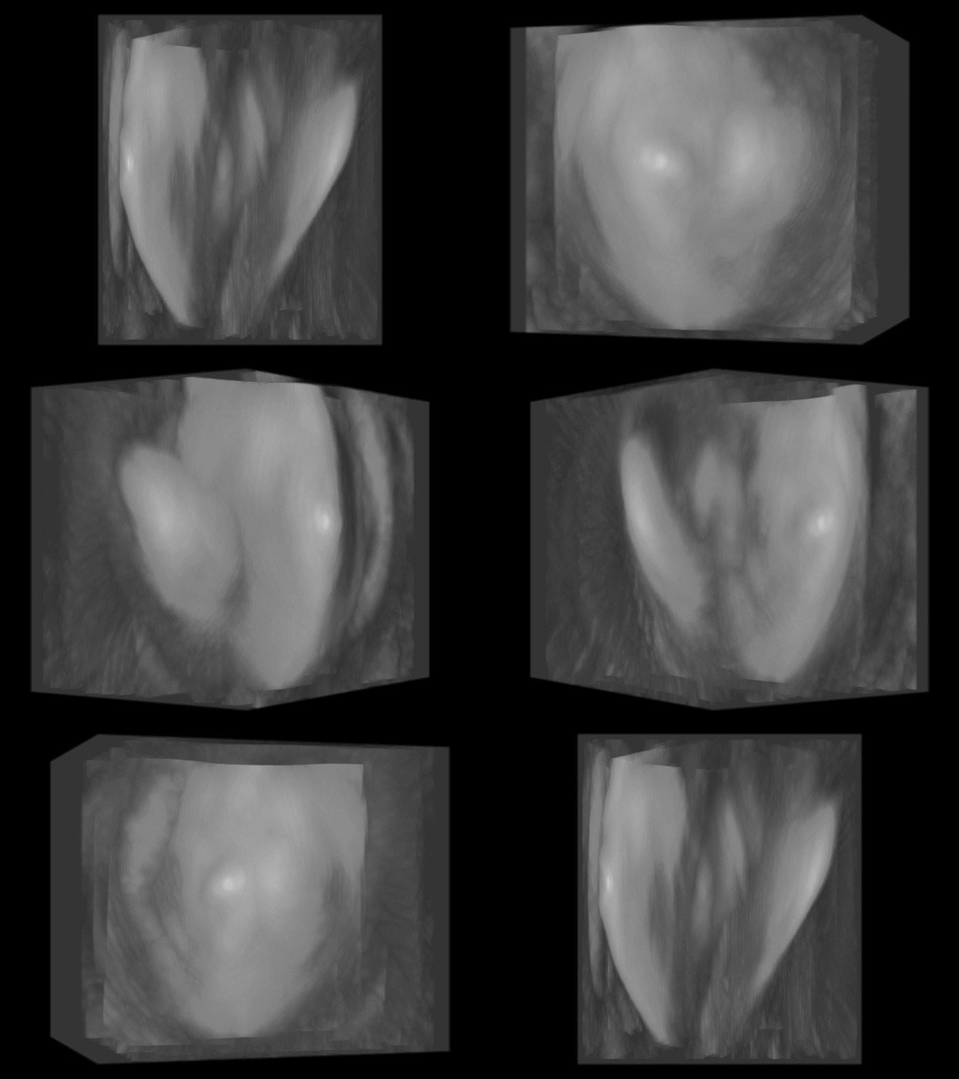
Animation einer 3D-Rekonstruktion eines Mäuseherzens mittels Optoakustischer Tomographie.
Optical Mapping
Im Gegensatz zur vielversprechenden optoakustischen Bildgebung, ist das Optical Mapping eine etablierte Methode, die es ermöglicht, Veränderungen der Zelleigenschaften (wie Membranspannung und Ionenkonzentration) mit hoher räumlicher und zeitlicher Auflösung zu erfassen, allerdings mit der Einschränkung, diese nur nahe der Oberfläche des Gewebes detektieren zu können. Wir haben ein Panoramabildgebungssystem entwickelt, mit dem wir mithilfe von Spiegeln die Oberfläche ganzer Herzen mit einer einzigen Kamera abbilden können. Dieses System wird derzeit weiterentwickelt, um die Korrelation der elektrischen Erregungsleitung über das Epikard mit der zugrunde liegenden Gewebestruktur mithilfe von Lichtblatt-Mikroskopie zu ermöglichen. Das Optical Mapping kommt in einer Reihe von Projekten zum Einsatz, unter anderem zur Untersuchung der Mechanismen der Arrhythmogenese bei Ischämie/Reperfusionsschädigung, sowie bei Bluthochdruck-induziertem kardialen Gewebeumbau.
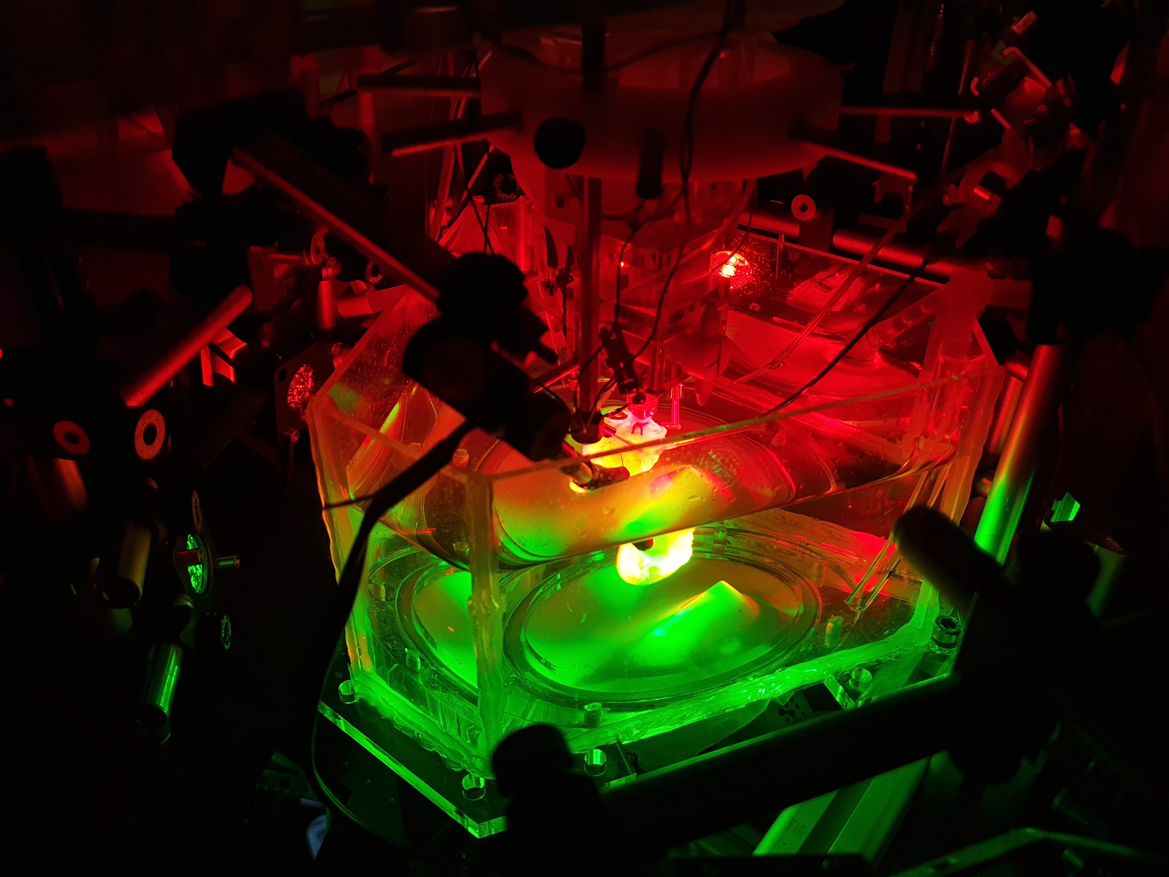
Panoramabildgebungssystem zur optischen Abbildung von Membran- spannung und Kalzium bei gleichzeitiger Messung der Mechanik mit Ultraschall.
Kardiale Stimulation
Wir können das Optical Mapping auch mit Werkzeugen zur optischen Kontrolle von Zellfunktionen kombinieren, z.B. via Licht-aktivierten Ionenpumpen und Kanälen. Solch ein kombinierter Aufbau ermöglicht somit vollkommen Licht-kontrollierte Untersuchungen kardiovaskulärer Funktionen von gesunden und krankheitsbedingt veränderten Proben. Ein Beispiel für solch eine Verwendung ist die optische Stimulation, bei der ein Herzschlag durch Bestrahlung mit Licht angeregt wird, anstatt durch einen elektrischen Strom (wie bei heutigen Herzschrittmachern üblich). Prinzipiell ist jedes Verfahren, das zu einem ausreichend großen Stromfluss in eine bestimmte Anzahl von Herzzellen führt, ausreichend, um einen Herzschlag zu beschleunigen. Interessanterweise können über dehnungs- aktivierte Kanäle im Herzen ähnliche Ströme hervorgerufen werden, d.h. ein Herzschlag kann durch bloßes Antippen beschleunigt werden. Dies ist besonders interessant für Notfallsituationen einer primären Asystole, wenn das Herz also nicht mehr schlägt und eine Herzmassage die beste verfügbare Option ist. Das erzeugbare Herzschlagvolumen ist jedoch relativ gering, so dass die Fähigkeit, das Herz durch einfaches Klopfen auf die Brust wieder zu beschleunigen, äußerst attraktiv erscheint. In ersten Experimenten konnte jedoch gezeigt werden, dass nach 50-100 Schlägen die mechanische Stimulierbarkeit abnimmt (Quinn et al., 2016). Um zu untersuchen, wie und warum dies geschieht, haben wir in Zusammenarbeit mit der Technischen Fakultät (IMTEK) ein System entwickelt, bei dem wir elektrische, mechanische und optische Stimulation an der exakt gleichen Stelle des Herzens anwenden und schnell zwischen den einzelnen Stimuli wechseln können.
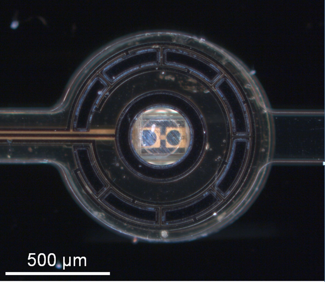
Spitze einer Optrodensonde bestehend aus einer 270 μm x 270 μm x 270 μm LED in der Mitte, umgeben von Elektroden.
- Giardini F, Olianti C, Marchal GA, Campos F, Romanelli V, Steyer J, Madl J, Piersanti R, Arecchi G, Vanaja IP, Biasci V, Rog-Zielinska EA, Nesi G, Loew LM, Cerbai E, Chelko SP, Regazzoni F, Loewe A, Bishop MJ, Mongillo M, Kohl P, Zaglia T, Zgierski-Johnston CM, Sacconi L. Correlative imaging integrates electrophysiology with three-dimensional murine heart reconstruction to reveal electrical coupling between cell types. Nat Cardiovasc Res 2025/4:1466-1486
- Mohr T, Schiffer M, Ramanujam D, Carls E, Zgierski-Johnston CM, Kok T, Niemann P, Geisen C, Kohl P, Engelhardt S, Fleischmann BK, Roell W. Efficient in vivo targeting of the myocardial scar using Moloney murine leukaemia virus complexed with nanoparticles. J Physiol 2025/604:1708-1735
- Chleilat E, Walz TP, Quinn TA, Kohl P, Zgierski-Johnston CM. Perivascular excitation tunnelling as a novel mechanism of cardiac reperfusion arrhythmias. bioRxiv 2023
- Fernández MC, Wülfers EM, Madl J, Perez Feliz S, Kohl P, Zgierski-Johnston CM, Schneider-Warme F. 3D structure of fibroblasts and macrophages in the healthy and cryo-ablated heart. bioRxiv 2023
- Becker C, Hardarson J, Hoelzer A, Schulz T, Reichl C, Burton NC, Schuler T, Kohl P, Zgierski-Johnston C. Evaluation of cervical lymph nodes using multispectral optoacoustic tomography: a proof-of-concept study. Eur Arch Otorhinolaryngol 2023/280:4657-64
- Johnston CM, Rog-Zielinska EA, Wülfers EM, Houwaart T, Siedlecka U, Naumann A, Nitschke R, Knöpfel T, Kohl P, Schneider-Warme F. Optogenetic targeting of cardiac myocytes and non-myocytes: Tools, challenges and utility. Prog Biophys Mol Biol 2017/130:140-149
- Sassu E, Tumlinson G, Stefanovska D, Fernández MC, Iaconianni P, Madl J, Brennan TA, Koch M, Cameron BA, Preissl S, Ravens U, Schneider-Warme F, Kohl P, Zgierski-Johnston CM, Hortells L. Age-related structural and functional changes of the intracardiac nervous system. J Mol Cell Cardiol. 2023 187:1-14
- Lewetag RD, Nimani S, Alerni N, Hornyik T, Jacobi SF, Moss R, Menza M, Pilia N, Walz TP, HajiRassouliha A, Perez-Feliz S, Zehender M, Seemann G, Zgierski-Johnston CM, Lopez R, Odening KE. Mechano-electrical interactions and heterogeneities in wild-type and drug-induced long QT syndrome rabbits. J Physiol. 2023/Epub ahead of print.
- Kohl P, Zgierski-Johnston CM. Assessment of Tissue Viability by Functional Imaging of Membrane Potential. Methods Mol Biol. 2023/2644:423-34
- Han B, Trew M, Zgierski-Johnston CM. Cardiac Conduction Velocity, Remodeling and Arrhythmogenesis. Cells 2021/10:2923
- Zgierski-Johnston CM, Ayub S, Fernández MC, Rog-Zielinska EA, Barz F, Paul O, Kohl P, Ruther P. Cardiac pacing using transmural multi-LED probes in channelrhodopsin-expressing mouse hearts. Prog Biophys Mol Biol 2020/154:51-61
Verfügbare Studentische Projekte
Die Abteilung Bioinstrumentation / Experimentelle Bildgebung sucht motivierte Studentinnen und Studenten für die folgenden Projekte . Sollten wir Ihr Interesse geweckt haben, so senden Sie bitte Ihren Lebenslauf (gern auf Englisch) an callum.johnston@uniklinik-freiburg.de.
Myocardial infarction remains a leading cause of mortality worldwide. Rapid reperfusion of occluded coronary arteries is essential to salvage ischaemic myocardium but paradoxically causes ischaemia-reperfusion arrhythmias (IRA). In previous work, we identified a new type of ischemia reperfusion arrhythmia occurring in the myocardium along the main branch of the reperfused vessel (“perivascular excitation tunnelling”, PVET) and showed that a 2-step reperfusion method could prevent them. We are now testing the efficacy of this reperfusion method as well a novel coronary catheter to deliver it in a translationally relevant pig model.
Background: Following an ischaemia-reperfusion event, arrhythmia can occur at the border zone or around the main trunk of the reperfused coronary vessel. Using optical mapping, we can assess the occurrence of these arrhythmias, and test the efficacy of our novel reperfusion method to prevent them.
Problem: Arrhythmia characterisation, interpretation and quantification are subjected to individual biases and approximations.
Aim: Develop a systematic workflow based on optical mapping data to identify and characterise arrhythmias following ischaemia-reperfusion. Ideally, this would be a semi-automated approach, but identification of key features would also be plausible.
Skills the candidate will learn:
Surgical preparation and help during the surgery of a large mammal;
Optical mapping of large mammal explanted heart;
Understanding of cardiac electrophysiology;
Analysis of optical mapping recordings;
Software and analysis workflow development.
Background: Perivascular excitation tunnelling (PVET) occurs around the main trunk of the reperfused vessel and can potentially lead to deadly re-entrant circuits. To assess its clinical relevance and assess whether it also occurs in patients, we need to identify a specific ECG signature pattern.
Problem: Analysis software inter-operability and current limited ECG spatial resolution make the identification of a specific ECG signature pattern to identify PVET difficult.
Aim: Improve the current analysis and experimental workflow to better combine optical maps and ECG traces to identify a PVET signature pattern that could be used in clinics.
Skills the candidate will learn:
Surgical preparation and help during the surgery of a large mammal;
Optical mapping of large mammal explanted heart;
Analysis of optical mapping recordings;
Understanding of cardiac electrophysiology;
Analysis and technical understanding of ECG data acquisition to improve its quality.
Background: When Ischaemia-reperfusion arrhythmia (IRA) occur in hospital, patients are “simply” defibrillated, but the presence of IRA is linked to poorer ventricular function recovery and higher short-term mortality.
Problem: There are currently no guidelines for identifying patients at risk of developing IRA, depending on ischaemic “structural parameters” e.g. size, location, transmurallity, number of occluded vessels amongst others.
Aim: Record ex vivo and develop in sillico different coronary architecture meshes to simulate various ischaemic contexts and identify which are the most arrhythmogenic, using computational simulations.
Subproject 1. MSc cand. /Dr. med. cand. project
MRI data acquisition and analysis, correlation of IRA occurrence (ECG based) with ischaemia “structural parameters” and histological assessment of the tissue viability in the area at risk (ischaemic area).
Skills the candidate will learn:
Surgical preparation and help during the surgery of a large mammal;
Ex vivo Langendorff perfusion experiments in a Magnetic Resonance Imaging (MRI) scanner;
Ex vivo ECG interpretation and analysis;
Analysis and 3D segmentation of gadolinium enhanced MRI images;
Histology assessment of tissue viability using TTC.
Subproject 2. Bioengineering project
Utilisation of high-resolution MRI segmented data to generate anatomically realistic meshes and assess the arrhythmogenic potential of ischaemia of various “structural parameters”
Skills the candidate will learn:
Segmentation of MR images;
Anatomically realistic mesh generation for computational modelling simulations;
Understanding of cellular and tissue electrophysiology principles,
Understanding of the methods to record cellular and tissue electrophysiology and their interpretation;
Computational simulations of cardiac electrophysiology.
Chicken embryos are an emerging 3R compliant model for examining cardiac physiology. Despite increasing attention by the scientific community, cardiac electrophysiology of the model remains incompletely characterised, limiting its translational potential.
Background: Chicken embryos are an emerging 3R compliant model for examining cardiac physiology. Despite increasing attention by the scientific community, cardiac electrophysiology of the model remains incompletely characterised, limiting its translational potential.
Problem: It is currently unclear to what extent the chicken embryo heart ion channel expression, function and response to drugs is similar to human.
Aim: The aim of this project is to characterise chicken embryo cardiac electrophysiology, and assess its comparability to human using various techniques (optical mapping, patch-clamp, tissue-slices).
Skills the candidate will learn:
Surgical preparation of small non-mammal heart;
Understanding of cellular and tissue electrophysiology;
Optical mapping recording and analysis;
Cardiomyocyte isolation;
Patch-clamp on freshly isolated cardiomyocytes.
Background: Chicken embryos are an emerging 3R compliant model for examining cardiac physiology. Despite increasing attention by the scientific community, cardiac electrophysiology of the model remains incompletely characterised, limiting its translational potential.
Problem: Due to the limited amount of data available on the chicken embryo heart, no computational model is currently available.
Aim: The aim of this project is to develop a computational model built upon the recordings obtained using various methods (optical mapping, patch-clamp, sharp-microelectrode) in our laboratory as well as pre-existing literature. This will be used to accurately simulate the electrophysiology of the model, and predict arrhythmogenic conditions.
Skills the candidate will learn:
Building of a novel computational model to accurately simulate cellular and tissue electrophysiology of an emerging study model;
Understanding of cellular and tissue electrophysiology principles,
Understanding of the methods to record cellular model and tissue electrophysiology and their interpretation;
Computational simulations of cardiac electrophysiology using OpenCARP.
Establishment of a tissue clearing and staining workflow for cardiac tissue.
Dr. med. cand. project
Background: Following cardiac injury, cell and protein spatial distribution and expression change to adapt to hostile environment, which results in functional modification of their behaviour.
Problem: Traditionally, 3D histological assessment requires the thin slicing (10 µm thick) of the preparation and complex computational approaches to realign and stack the slices, which is not compatible with centimetre-thick preparations.
Aim: Establish an experimental workflow for the clearing of millimetre to centimetre-thick tissues for 3D histological assessment and reconstruction.
Skills the candidate will learn:
Preparation for and assistance during the surgery on a large mammal;
Tissue clearing of small non-mammal (chicken embryo) as well as small (mouse) and large mammal (rabbit and pig heart) hearts;
Sample preparation for histological imaging;
Microscopic imaging and analysis of protein distribution.
Projecting Electrophysiological Data onto 3D Surface Reconstruction of Whole Hearts.
Bioengineering project
Background: Optical mapping allows for the tracking of important electrophysiological changes across the surface of the heart, including action potential duration and conduction velocity, though most methods use a single 2D perspective of one side of the heart, which is neither flat nor homogeneous. Panoramic vision systems acquire images of the whole heart surface, allowing for a more comprehensive and spatially accurate understanding of these electrophysiological changes.
Problem: The images used for 3D reconstruction of the heart surface also contain electrophysiological data, but aligning the two datasets (structural and electrophysiological) across multiple views is challenging. For contracting hearts, motion artifacts and time-varying geometry introduce further complication for data alignment.
Aim: Establish a rules-based workflow to align structural and functional electrophysiological data in 3D without manual landmark selection.
Skills the candidate will learn:
Ray tracing and gaussian splatting in panoramic imaging systems;
Cutting-edge 3D reconstruction and motion tracking methodologies;
Modelling of emitted light from non-homogeneous, contracting biological tissue;
Deriving electrophysiological properties from optical mapping data in 3D;
Working with data from multiple animal models (mouse, chicken, and rabbit).
Coronary Flow Dynamics during Pharmacological Activation of Piezo1.
MSc cand. /Dr. med. cand. project
Background: Piezo1 is a mechanosensor that is important for a range of physiological and pathological functions. In the heart, Piezo1 has been linked to fibrosis and hypertrophy as well as to acute changes in electrophysiology.
Problem: We have recently identified that pharmacological activating of Piezo1 with Yoda1 induces significant reduction in the coronary flow of Langendorff-perfused whole hearts. The mechanism and cell types involved, however remains uncertain.
Aim of the project: Assess different mechanisms by which Piezo1 may regulate vascular tone.
Skills developed:
Langendorff-perfusion of murine hearts;
Optical mapping of cardiac electrophysiology and calcium;
Isolation and patch clamp of single cells;
Assessment of changes in cardiac haemodynamics and electrocardiography during drug perfusion.
Team

Dr. Callum Zgierski-Johnston
Arbeitsgruppenleiter

Leonardo Sacconi, PhD
Senior Scientist

Dr. Francesco Giardini

Dr. Tony Rubio
E-Mail: tony.rubio@uniklinik-freiburg.de

Thomas Kok
E-Mail: thomas.kok@uniklinik-freiburg.de

Collin Snitchler

E-Mail: bo.han@uniklinik-freiburg.de

Anna-Lena Herm

Jana Ebeling

Gyuwon Lee
E-Mail: gyuwon.lee@uniklinik-freiburg.de