Mechanobiology
The Mechanobiology Group studies the signalling processes related to mechanical forces that are omnipresent and constantly changing in the heart. They shape life, acting either as crucial drivers for a wide range of physiological processes, or as potential threats to which cells have to adapt to survive in pathological conditions. How cells sense their environment and adapt their function to changes in mechanical states is the key question driving this group. Molecular mechanisms underlying this ability are poorly understood and we focus on mechano-sensitive channels, key players in Mechano-Electric Feedback (MEF).
Molecular identity and functions of cardiac mechano-sensitive channels
To unravel functions of these mechano-sensors, we combine the patch-clamp technique with equipment that allows:
- acute mechanical stimulation of minute membrane areas, such as by high speed pressure clamp, to stretch patches of membrane inside a recording pipette;
- chronic mechanical stimulation, such as using the Flexcell® system, to expose cells in culture to changes that mimic normal or diseased cardiovascular Function.
Using genetic tools, and pharmacological interventions we target individual channel candidates, and test their roles in cells ranging from cardiac muscle cells to connective tissue and heart valve endothelium.
Fig.1: Glass capillaries that will be sealed to the cell plasma membrane to record ion channel activity. Patch-clamp is one of the very few techniques that allow one to observe conformational changes of a single protein in real time.
Axial and radial forces in isolated cardiomyocytes
Combining our Carbon Fibre technique (see 4D Imaging page) and Atomic Force Microscopy (from AFM Workshop, Fig. 2) we can record both axial and radial forces from single isolated cardiomyocytes in different loading states. We use this to study acute mechanical effects on cell mechanical properties and, by combining this with fluorescent imaging and patch clamp investigations, we explore ionic mechanisms underlying observed behaviour.
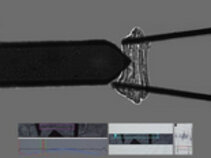
Fig.2: A ventricular cardiomyocyte stretched by two carbon fibres (right) under the cantilever of an atomic force microscope (left). Carbon fibres allow one to prescribe and record axial force development, whilst the AFM cantilever records radial forces. Cells are paced, perfused and temperature-controlled. (Source: AFM Workshop)
In the long term, whilst modifying the loading state of single isolated cardiomyocytes, we plan to better understand the distribution of mechanical forces in the cytoskeleton and at the plasma membrane level. To do so, we will use fluorescent tension probes and fluorescent curvature-sensitive probes. This is anticipated to allow us to understand where and how changes in membrane curvature / cytoskeletal tension occur, and what impact they have on cellular mechano-sensing.
- Künzel SR [...] Dudek S, Emig R, Ravens U, Rog-Zielinska EA, Peyronnet R, El-Armouche A. Modeling atrial fibrosis in vitro-Generation and characterization of a novel human atrial fibroblast cell line. FEBS Open Bio 2020/10:1210-1218
- Darkow E, Rog-Zielinska EA, Madl J [...] Kohl P, Ravens U, Römer W, Peyronnet R. The Lectin LecA Sensitizes the Human Stretch-Activated Channel TREK-1 but Not Piezo1 and Binds Selectively to Cardiac Non-myocytes. Front Physiol 2020/11:457
- Emig R, Zgierski-Johnston CM, Beyersdorf F, Rylski B, Ravens U, Weber W, Kohl P, Hörner M, Peyronnet R. Human Atrial Fibroblast Adaptation to Heterogeneities in Substrate Stiffness. Front Physiol 2020/10:1526
- Peyronnet R, Ravens U. Atria-selective antiarrhythmic drugs in need of alliance partners. Pharmacol Res 2019/145:104262
- Schmitt R, Tscheuschler A [...] Peyronnet R, Kari FA. A potential key mechanism in ascending aortic aneurysm development: Detection of a linear relationship between MMP-14/TIMP-2 ratio and active MMP-2. PLoS One 2019/14:e0212859
- Odening KE, Bodi I [...] Perez-Feliz S, Fürniss H, Mettke L, Michaelides K [...] Peyronnet R [...] Bode C, Jolivet G, Brunner M. Transgenic short-QT syndrome 1 rabbits mimic the human disease phenotype with QT/APD shortening in the atria and ventricles and increased VT/VF inducibility. Eur Heart J 2019/40:842-853
- Klesen A, Jakob D, Emig R, Kohl P, Ravens U, Peyronnet R. Cardiac fibroblasts: Active players in (atrial) electrophysiology? Herzschrittmacherther Elektrophysiol 2018/29:62-69
- Rog-Zielinska EA and Peyronnet R. Cardiac mechanics and electrics: It takes two to tango. Prog Biophys Mol Biol 2017/130:121-123
- Peyronnet R, Bollensdorff C, Capel RA, Rog-Zielinska EA, Woods CE, Charo DN, Lookin O, Fajardo G, Ho M, Quertermous T, Ashley EA, Kohl P. Load-dependent effects of apelin on murine cardiomyocytes. Prog Biophys Mol Biol 2017/130:333-343
- Burton RAB, Rog-Zielinska EA, Corbett AD, Peyronnet R, Bodi I, Fink M, Sheldon J, Hoenger A, Calaghan SC, Bub G, Kohl P. Caveolae in Rabbit Ventricular Myocytes: Distribution and Dynamic Diminution after Cell Isolation. Biophys J 2017/113:1047-1059
- Decher N, Ortiz-Bonnin B [...] Seemann G, Peyronnet R, Zumhagen S, Bustos D, Kockskämper J, Kohl P [...] Schulze-Bahr E. Sodium permeable and "hypersensitive" TREK-1 channels cause ventricular tachycardia. EMBO Mol Med 2017/9:403-414
- Peyronnet R, Nerbonne J, Kohl P. Cardiac mechano-gated ion channels and arrhythmias. Circ Res 2016/118:311-329
- Lohezic M, Teh I, Bollensdorff C, Peyronnet R, Hales PW, Grau V, Kohl P & Schneider JE. Interrogation of living myocardium in multiple static deformation states with diffusion tensor and diffusion spectrum imaging. Prog Biophys Mol Biol 2014/115:213-2259
- Reed A, Kohl P, Peyronnet R. Molecular candidates for cardiac stretch-activated ion channels. Glob Cardiol Sci Pract 2014/2014:9-25
- Iribe G, Ward CW, Camelliti P, Bollensdorff C, Mason F, Burton RA, Garny A, Morphew MK, Hoenger A, Lederer WJ & Kohl P. Axial stretch of rat single ventricular cardiomyocytes causes an acute and transient increase in Ca2+ spark rate. Circ Res 2009/104:787-795
Team

Dr. Rémi Peyronnet
Head of section

Prof. Ursula Ravens
Senior professor

Arthur Boileve, PhD

Anthony Coté

Marine Devaux

Hanna Hemeling

Adrian-Yarema Holota

Minou Neidlein
