

Bioinstrumentation / Experimental Imaging
The Bioinstrumentation / Experimental Imaging Group develops and applies specialised equipment for cardiovascular research. This is required as often our questions cannot be addressed with off-the-shelf solutions. To this end, we have facilities for prototyping of mechanical and electrical components, and expertise in sensors, imaging, signal processing, control systems and user-interfacing.
The resulting systems are used to examine cardiac physiology with a particular focus on electrophysiology and the mechanisms underlying ventricular arrhythmias. By understanding the mechanisms of arrhythmias, we can then investigate possible prevention and treatment options including the use of drugs, targeted genetic modification and medical device development.
Optoacoustic imaging
Currently there is no way to directly image the electrical activity in the heart in 3D in a tissue non-destructive manner. Optoacoustic imaging is a method where laser pulses are used to excite tissue, resulting in thermoelastic expansion of chromophores with the tissue and subsequently to generation of a pressure wave that can be recorded by ultrasound. By using tomographic techniques the exact location of the chromophores within the tissue can be identified. Optoacoustic imaging, by combining the advantages of fluorescent imaging with the penetration depth of ultrasound, holds the potential to enable measurement of cardiac electrophysiology in 3D.
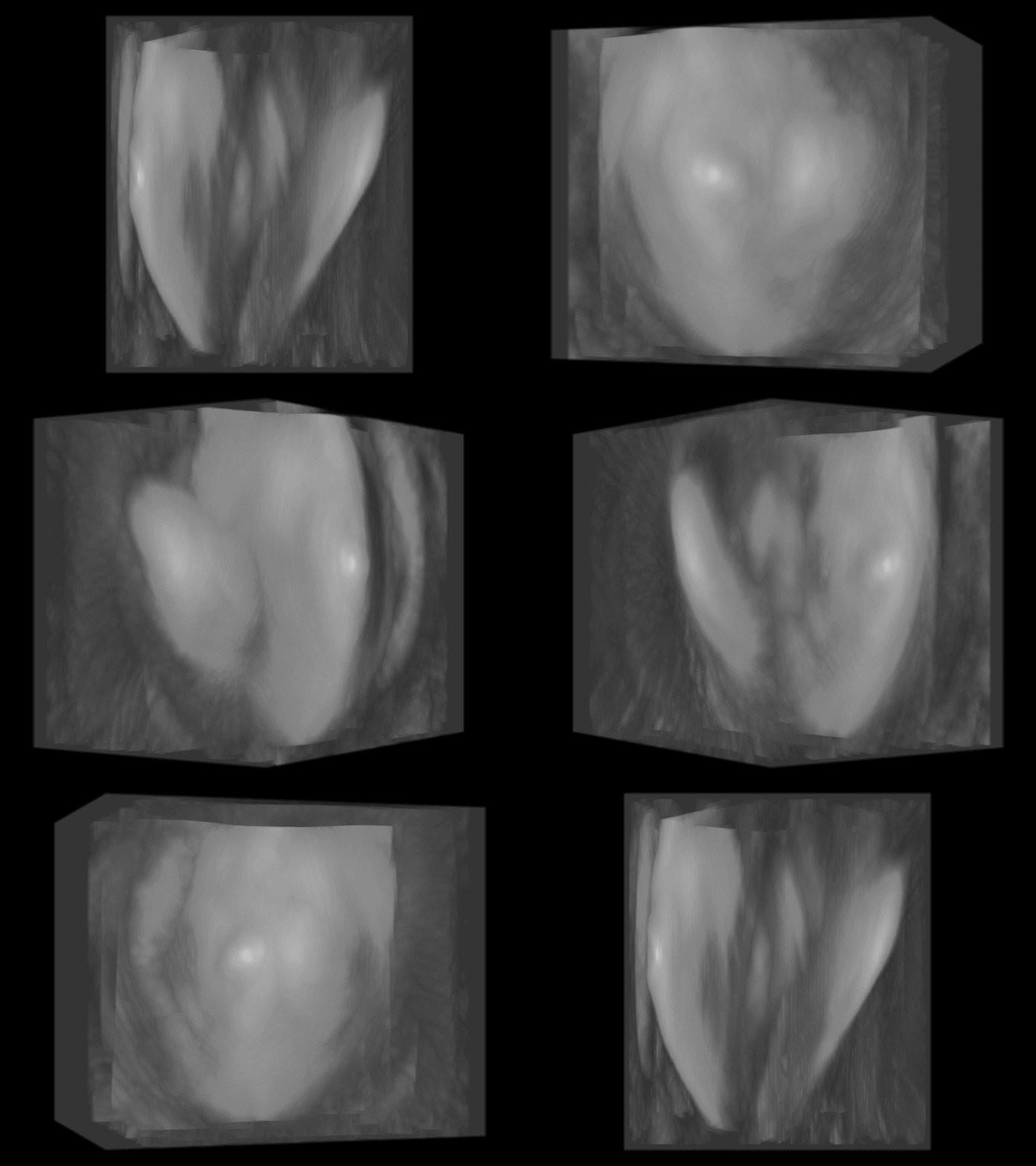
Animation of a 3D reconstruction of a mouse heart imaged using optoacoustic tomography.
Optical Mapping
While optoacoustic imaging holds great promise to measure cardiac electrophysiology, optical mapping is a relatively mature technique enabling the measurement of changes in cell function (such as membrane voltage and ion concentration) at high spatial and temporal resolution albeit with the limitation of only measuring these at the surface or near-surface of the tissue. We have developed panoramic imaging systems, which allow us to image the epicardial surface of whole hearts. This is being further developed to allow correlation of electrical conduction across the epicardium with the underlying tissue structure using advanced light-sheet based imaging methods. The optical mapping system is used in a number of projects including investigating the mechanisms underlying arrhythmia formation in ischaemia/reperfusion injury and upon pressure-overload induced remodelling.
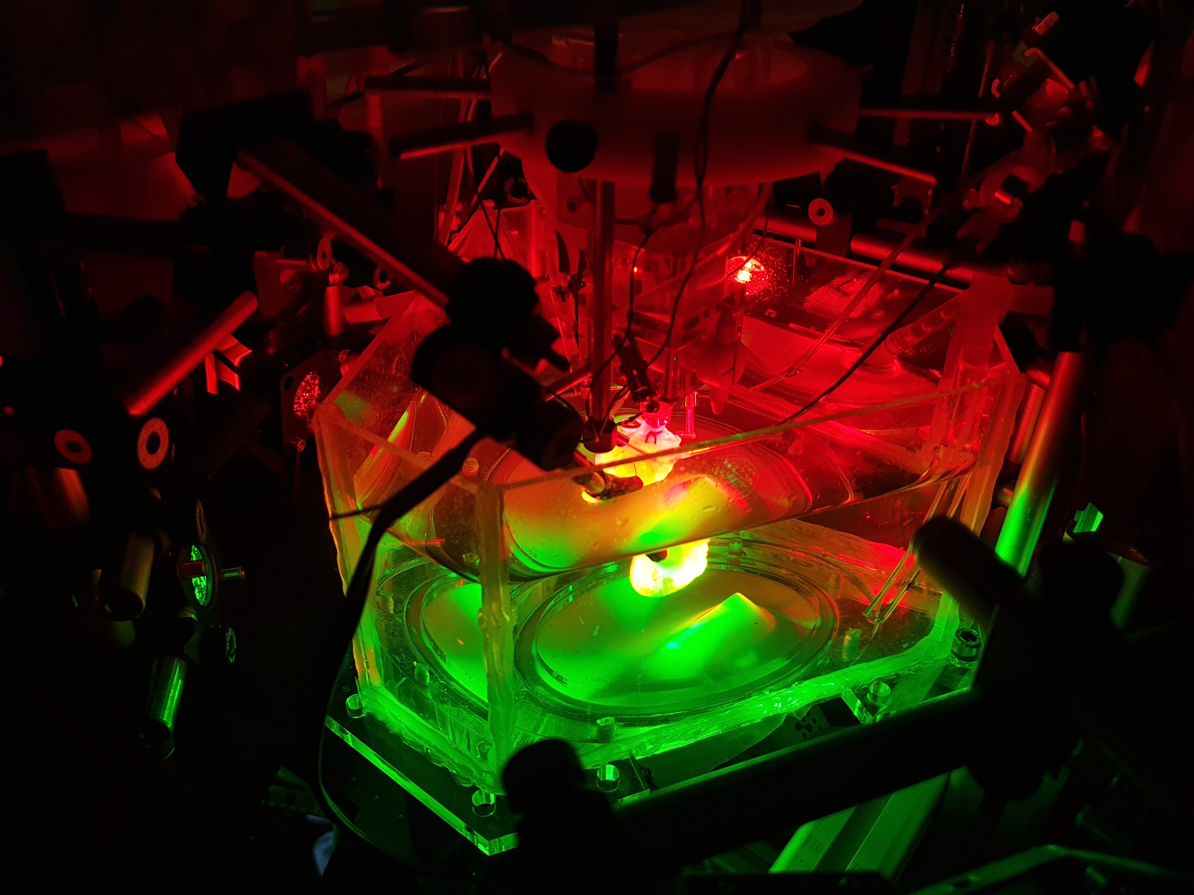
Panoramic imaging system for optical mapping of membrane voltage and calcium while simultaneously measuring mechanics with ultrasound.
Cardiac pacing
We can also combine optical mapping with tools for optical control of function, via light-activated ion pumps and channels. These tools open up doors towards fully light-controlled studies of cardiovascular function in both health and disease. An example of their use is for optical pacing, where the heart can be paced by shining light on it instead of passing an electric current through it. In principle any method resulting in a sufficiently large flow of current into a set number of cardiac cells is sufficient to pace the heart, thus tapping the heart can also pace it via stretch-activated channels. This mechanical pacing is potentially interesting in the emergency setting of primary asystole where the heart is not beating and there is no specialized care nearby allowing rapid electrical pacing. In this setting, CPR is the best option, however the output from contractions is relatively low, thus the ability to pace the heart by just tapping on the chest is attractive. Unfortunately, mechanical pacing is not sustainable with initial experiments demonstrating a loss in the ability to pace after 50 to 100 beats (Quinn et al., 2016). To investigate how and why this occurs we have, in collaboration with the technical faculty (IMTEK), developed a system where we can use electrical, mechanical and optical pacing at the same location and rapidly switch between techniques to better understand the limitations of such pacing.
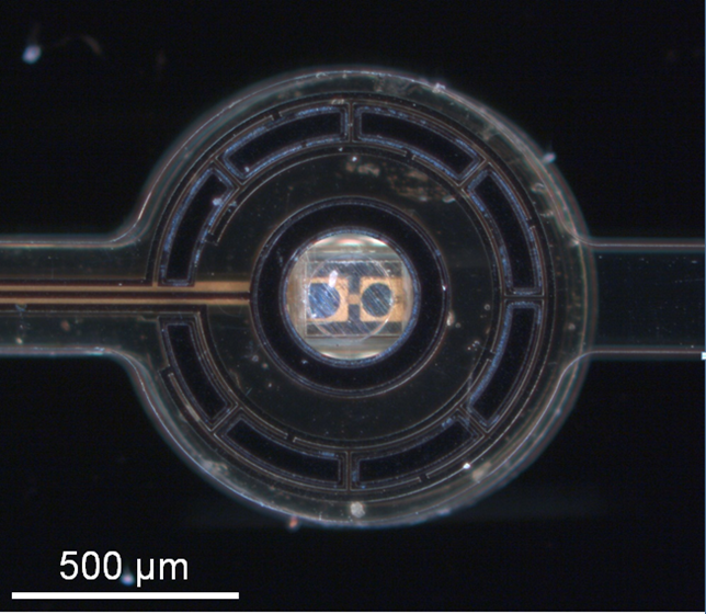
Tip of an optrode-probe comprising a 270 μm x 270 μm LED in the centre with surrounding electrodes.
- Giardini F, Olianti C, Marchal GA, Campos F, Romanelli V, Steyer J, Madl J, Piersanti R, Arecchi G, Vanaja IP, Biasci V, Rog-Zielinska EA, Nesi G, Loew LM, Cerbai E, Chelko SP, Regazzoni F, Loewe A, Bishop MJ, Mongillo M, Kohl P, Zaglia T, Zgierski-Johnston CM, Sacconi L. Correlative imaging integrates electrophysiology with three-dimensional murine heart reconstruction to reveal electrical coupling between cell types. Nat Cardiovasc Res 2025/4:1466-1486
- Mohr T, Schiffer M, Ramanujam D, Carls E, Zgierski-Johnston CM, Kok T, Niemann P, Geisen C, Kohl P, Engelhardt S, Fleischmann BK, Roell W. Efficient in vivo targeting of the myocardial scar using Moloney murine leukaemia virus complexed with nanoparticles. J Physiol 2025/604:1708-1735
- Chleilat E, Walz TP, Quinn TA, Kohl P, Zgierski-Johnston CM. Perivascular excitation tunnelling as a novel mechanism of cardiac reperfusion arrhythmias. bioRxiv 2023
- Fernández MC, Wülfers EM, Madl J, Perez Feliz S, Kohl P, Zgierski-Johnston CM, Schneider-Warme F. 3D structure of fibroblasts and macrophages in the healthy and cryo-ablated heart. bioRxiv 2023
- Becker C, Hardarson J, Hoelzer A, Schulz T, Reichl C, Burton NC, Schuler T, Kohl P, Zgierski-Johnston C. Evaluation of cervical lymph nodes using multispectral optoacoustic tomography: a proof-of-concept study. Eur Arch Otorhinolaryngol 2023/280:4657-64
- Johnston CM, Rog-Zielinska EA, Wülfers EM, Houwaart T, Siedlecka U, Naumann A, Nitschke R, Knöpfel T, Kohl P, Schneider-Warme F. Optogenetic targeting of cardiac myocytes and non-myocytes: Tools, challenges and utility. Prog Biophys Mol Biol 2017/130:140-149
- Sassu E, Tumlinson G, Stefanovska D, Fernández MC, Iaconianni P, Madl J, Brennan TA, Koch M, Cameron BA, Preissl S, Ravens U, Schneider-Warme F, Kohl P, Zgierski-Johnston CM, Hortells L. Age-related structural and functional changes of the intracardiac nervous system. J Mol Cell Cardiol. 2023 187:1-14
- Lewetag RD, Nimani S, Alerni N, Hornyik T, Jacobi SF, Moss R, Menza M, Pilia N, Walz TP, HajiRassouliha A, Perez-Feliz S, Zehender M, Seemann G, Zgierski-Johnston CM, Lopez R, Odening KE. Mechano-electrical interactions and heterogeneities in wild-type and drug-induced long QT syndrome rabbits. J Physiol. 2023/Epub ahead of print.
- Kohl P, Zgierski-Johnston CM. Assessment of Tissue Viability by Functional Imaging of Membrane Potential. Methods Mol Biol. 2023/2644:423-34
- Han B, Trew M, Zgierski-Johnston CM. Cardiac Conduction Velocity, Remodeling and Arrhythmogenesis. Cells 2021/10:2923
- Zgierski-Johnston CM, Ayub S, Fernández MC, Rog-Zielinska EA, Barz F, Paul O, Kohl P, Ruther P. Cardiac pacing using transmural multi-LED probes in channelrhodopsin-expressing mouse hearts. Prog Biophys Mol Biol 2020/154:51-61
Available Projects
The Bioinstrumentation / Experimental Imaging group is looking for interested students for the following projects. If you’re interested or want to hear more, please send a CV to callum.johnston@uniklinik-freiburg.de.
Myocardial infarction remains a leading cause of mortality worldwide. Rapid reperfusion of occluded coronary arteries is essential to salvage ischaemic myocardium but paradoxically causes ischaemia-reperfusion arrhythmias (IRA). In previous work, we identified a new type of ischemia reperfusion arrhythmia occurring in the myocardium along the main branch of the reperfused vessel (“perivascular excitation tunnelling”, PVET) and showed that a 2-step reperfusion method could prevent them. We are now testing the efficacy of this reperfusion method as well a novel coronary catheter to deliver it in a translationally relevant pig model.
Background: Following an ischaemia-reperfusion event, arrhythmia can occur at the border zone or around the main trunk of the reperfused coronary vessel. Using optical mapping, we can assess the occurrence of these arrhythmias, and test the efficacy of our novel reperfusion method to prevent them.
Problem: Arrhythmia characterisation, interpretation and quantification are subjected to individual biases and approximations.
Aim: Develop a systematic workflow based on optical mapping data to identify and characterise arrhythmias following ischaemia-reperfusion. Ideally, this would be a semi-automated approach, but identification of key features would also be plausible.
Skills the candidate will learn:
Surgical preparation and help during the surgery of a large mammal;
Optical mapping of large mammal explanted heart;
Understanding of cardiac electrophysiology;
Analysis of optical mapping recordings;
Software and analysis workflow development.
Background: Perivascular excitation tunnelling (PVET) occurs around the main trunk of the reperfused vessel and can potentially lead to deadly re-entrant circuits. To assess its clinical relevance and assess whether it also occurs in patients, we need to identify a specific ECG signature pattern.
Problem: Analysis software inter-operability and current limited ECG spatial resolution make the identification of a specific ECG signature pattern to identify PVET difficult.
Aim: Improve the current analysis and experimental workflow to better combine optical maps and ECG traces to identify a PVET signature pattern that could be used in clinics.
Skills the candidate will learn:
Surgical preparation and help during the surgery of a large mammal;
Optical mapping of large mammal explanted heart;
Analysis of optical mapping recordings;
Understanding of cardiac electrophysiology;
Analysis and technical understanding of ECG data acquisition to improve its quality.
Background: When Ischaemia-reperfusion arrhythmia (IRA) occur in hospital, patients are “simply” defibrillated, but the presence of IRA is linked to poorer ventricular function recovery and higher short-term mortality.
Problem: There are currently no guidelines for identifying patients at risk of developing IRA, depending on ischaemic “structural parameters” e.g. size, location, transmurallity, number of occluded vessels amongst others.
Aim: Record ex vivo and develop in sillico different coronary architecture meshes to simulate various ischaemic contexts and identify which are the most arrhythmogenic, using computational simulations.
Subproject 1. MSc cand. /Dr. med. cand. project
MRI data acquisition and analysis, correlation of IRA occurrence (ECG based) with ischaemia “structural parameters” and histological assessment of the tissue viability in the area at risk (ischaemic area).
Skills the candidate will learn:
Surgical preparation and help during the surgery of a large mammal;
Ex vivo Langendorff perfusion experiments in a Magnetic Resonance Imaging (MRI) scanner;
Ex vivo ECG interpretation and analysis;
Analysis and 3D segmentation of gadolinium enhanced MRI images;
Histology assessment of tissue viability using TTC.
Subproject 2. Bioengineering project
Utilisation of high-resolution MRI segmented data to generate anatomically realistic meshes and assess the arrhythmogenic potential of ischaemia of various “structural parameters”
Skills the candidate will learn:
Segmentation of MR images;
Anatomically realistic mesh generation for computational modelling simulations;
Understanding of cellular and tissue electrophysiology principles,
Understanding of the methods to record cellular and tissue electrophysiology and their interpretation;
Computational simulations of cardiac electrophysiology.
Chicken embryos are an emerging 3R compliant model for examining cardiac physiology. Despite increasing attention by the scientific community, cardiac electrophysiology of the model remains incompletely characterised, limiting its translational potential.
Background: Chicken embryos are an emerging 3R compliant model for examining cardiac physiology. Despite increasing attention by the scientific community, cardiac electrophysiology of the model remains incompletely characterised, limiting its translational potential.
Problem: It is currently unclear to what extent the chicken embryo heart ion channel expression, function and response to drugs is similar to human.
Aim: The aim of this project is to characterise chicken embryo cardiac electrophysiology, and assess its comparability to human using various techniques (optical mapping, patch-clamp, tissue-slices).
Skills the candidate will learn:
Surgical preparation of small non-mammal heart;
Understanding of cellular and tissue electrophysiology;
Optical mapping recording and analysis;
Cardiomyocyte isolation;
Patch-clamp on freshly isolated cardiomyocytes.
Background: Chicken embryos are an emerging 3R compliant model for examining cardiac physiology. Despite increasing attention by the scientific community, cardiac electrophysiology of the model remains incompletely characterised, limiting its translational potential.
Problem: Due to the limited amount of data available on the chicken embryo heart, no computational model is currently available.
Aim: The aim of this project is to develop a computational model built upon the recordings obtained using various methods (optical mapping, patch-clamp, sharp-microelectrode) in our laboratory as well as pre-existing literature. This will be used to accurately simulate the electrophysiology of the model, and predict arrhythmogenic conditions.
Skills the candidate will learn:
Building of a novel computational model to accurately simulate cellular and tissue electrophysiology of an emerging study model;
Understanding of cellular and tissue electrophysiology principles,
Understanding of the methods to record cellular model and tissue electrophysiology and their interpretation;
Computational simulations of cardiac electrophysiology using OpenCARP.
Establishment of a tissue clearing and staining workflow for cardiac tissue.
Dr. med. cand. project
Background: Following cardiac injury, cell and protein spatial distribution and expression change to adapt to hostile environment, which results in functional modification of their behaviour.
Problem: Traditionally, 3D histological assessment requires the thin slicing (10 µm thick) of the preparation and complex computational approaches to realign and stack the slices, which is not compatible with centimetre-thick preparations.
Aim: Establish an experimental workflow for the clearing of millimetre to centimetre-thick tissues for 3D histological assessment and reconstruction.
Skills the candidate will learn:
Preparation for and assistance during the surgery on a large mammal;
Tissue clearing of small non-mammal (chicken embryo) as well as small (mouse) and large mammal (rabbit and pig heart) hearts;
Sample preparation for histological imaging;
Microscopic imaging and analysis of protein distribution.
Projecting Electrophysiological Data onto 3D Surface Reconstruction of Whole Hearts.
Bioengineering project
Background: Optical mapping allows for the tracking of important electrophysiological changes across the surface of the heart, including action potential duration and conduction velocity, though most methods use a single 2D perspective of one side of the heart, which is neither flat nor homogeneous. Panoramic vision systems acquire images of the whole heart surface, allowing for a more comprehensive and spatially accurate understanding of these electrophysiological changes.
Problem: The images used for 3D reconstruction of the heart surface also contain electrophysiological data, but aligning the two datasets (structural and electrophysiological) across multiple views is challenging. For contracting hearts, motion artifacts and time-varying geometry introduce further complication for data alignment.
Aim: Establish a rules-based workflow to align structural and functional electrophysiological data in 3D without manual landmark selection.
Skills the candidate will learn:
Ray tracing and gaussian splatting in panoramic imaging systems;
Cutting-edge 3D reconstruction and motion tracking methodologies;
Modelling of emitted light from non-homogeneous, contracting biological tissue;
Deriving electrophysiological properties from optical mapping data in 3D;
Working with data from multiple animal models (mouse, chicken, and rabbit).
Coronary Flow Dynamics during Pharmacological Activation of Piezo1.
MSc cand. /Dr. med. cand. project
Background: Piezo1 is a mechanosensor that is important for a range of physiological and pathological functions. In the heart, Piezo1 has been linked to fibrosis and hypertrophy as well as to acute changes in electrophysiology.
Problem: We have recently identified that pharmacological activating of Piezo1 with Yoda1 induces significant reduction in the coronary flow of Langendorff-perfused whole hearts. The mechanism and cell types involved, however remains uncertain.
Aim of the project: Assess different mechanisms by which Piezo1 may regulate vascular tone.
Skills developed:
Langendorff-perfusion of murine hearts;
Optical mapping of cardiac electrophysiology and calcium;
Isolation and patch clamp of single cells;
Assessment of changes in cardiac haemodynamics and electrocardiography during drug perfusion.
Team

Dr. Callum Zgierski-Johnston
Head of section

Leonardo Sacconi, PhD
Senior Scientist


Dr. Tony Rubio
E-Mail: tony.rubio@uniklinik-freiburg.de

Thomas Kok
E-Mail: thomas.kok@uniklinik-freiburg.de

Collin Snitchler

E-Mail: bo.han@uniklinik-freiburg.de

Anna-Lena Herm

Jana Ebeling