Clinical MRI
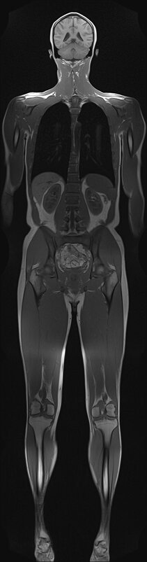
Unlike a number of diseases single MR examinations target only parts of the body not exceeding the length of the scanners field of view (FOV) of about 50cm. For the assessment of disease patterns with non localized findings we are interested in developing a technique for the seamless acquisition of any FOV by moving the patient continuously through the scanner during the measurement. Choosing the imaging plane perpendicular to the motion direction – similar to a CT scan – holds an appealing technical feature: Any moving 3D volume can be imaged within a small acquisition window of a single 2D slice. Our ultimate vision is to develop imaging methods for a new generation of MR scanners that look more like today’s ring-shaped CT scanners than the awesome tubes they used to be 20 years ago. For the successfull installation of extended FOV imaging in clinical practice we have ambitious goals regarding patient convenience and image quality of the examination. Therefore imaging techniques that can be performed during free breathing and provide isotropic high resolution data sets are major subject to our research activities. The evaluation of our developments in clinical studies has shown that they have great capability to make MR examinations more flexible and to simplify and speed up the imaging of large FOVs while providing highest image quality. And this not only on tomorrows short bore magnets but already on any today’s scanner.
Group Leader
Clinical Partners
Prof. Oliver Schäfer
Dr. Tobias Baumann
Dr. Julia Geiger

Dr. Ute Ludwig
Group Leader
Tel.: +49 761 270-38340
E-Mail: ute.ludwig@uniklinik-freiburg.de
University Medical Center Freiburg
Dept. of Radiology · Medical Physics
Killianstr. 5a
79106 Freiburg