
Spectral CT Imaging
Advanced CT Imaging and Visualization (ACTIV)
Hybrid Pixel Detector Imaging Lab
We develop compact, multimodal imaging systems based on hybrid pixel detector technology. Our work spans spectral CT, Compton and gamma imaging, and integrated imaging platforms for clinical, preclinical, and emerging applications.
Our central idea is to use a single detector technology paradigm to unify multiple imaging modalities:
- X-ray CT and radiography
- Spectral and photon-counting imaging
- Gamma and Compton imaging
- Hybrid systems combining CT and nuclear imaging
This enables fundamentally new system designs that are:
- Compact and mobile
- Quantitative and spectrally resolved
- Compatible with clinical and intraoperative environments
Core Research Directions
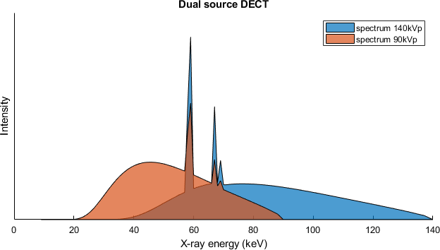
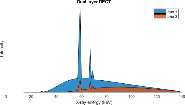
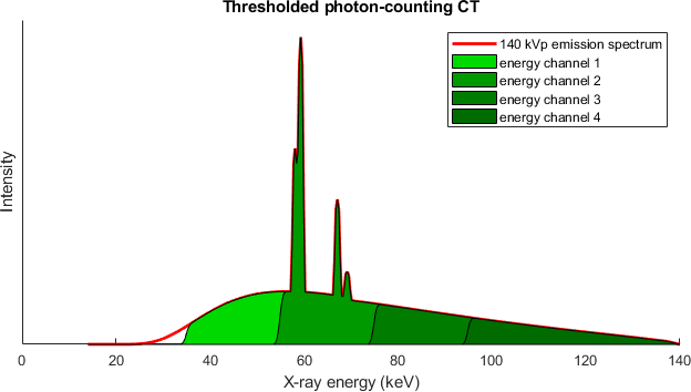
Spectral & Photon-Counting CT
Energy-resolved X-ray imaging for material decomposition, artifact reduction, and quantitative imaging.
Gamma & Compton Imaging Systems
Development of miniaturized Compton cameras and gamma imaging solutions for intraoperative and endoscopic use.
Multimodal Imaging Platforms
Integration of CT, gamma imaging, and MRI into unified acquisition and reconstruction frameworks.
Computational Imaging & Reconstruction
Physics-based and data-driven reconstruction methods for sparse, noisy, and heterogeneous data.
System Vision
Our research aims at breaking classical modality boundaries.
We develop imaging systems that:
- Combine anatomical and functional information in a single setup
- Operate with minimal infrastructure
- Enable real-time and intraoperative imaging
- Scale from preclinical systems to clinical translation
This includes:
- Compact hybrid CT/gamma systems
- Endoscopic and laparoscopic gamma imaging
- Mobile or infrastructure-independent imaging solutions
Applications
- Oncology and molecular imaging
- Surgical guidance and intraoperative imaging
- Dental and high-resolution imaging
- Preclinical and biomaterial research

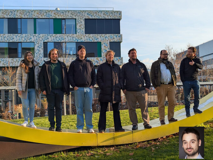
Dr. Martin Peter Pichotka
Scientific Lead of the Spectral CT Research Group
Tel.: +49 761 270-39220
E-Mail: martin.pichotka@uniklinik-freiburg.de
Medical Center – University of Freiburg
Dept. of Radiology · Medical Physics
Killianstr. 5a
79106 Freiburg