Applications
Micro-CT imaging
CT imaging
CT is a 3D imaging modality, based on the acquisition of a series of radiographs and their consecutive volumetric reconstruction. The result of a classic CT scan yields a volumetric representation of the patient / sample featuring grey value encoded CT numbers, which in turn represent the local material density multiplied by the convolution of the X-ray spectrum employed and the respective material’s energy dependent X-ray attenuation coefficient. Given a sufficiently homogeneous material composition and narrow X-ray spectrum, the respective gray values give an approximate measure of local material density.
Key characteristics in classic CT imaging are:
- Achievable volumetric resolution
- Contrast between constituent tissue types / materials
- Required patient dose / machine time
- Noise level within the volumetric image
- Presence and magnitude of imaging artefacts
These characteristics are influenced by a number of factors, including:
- Detector characteristics (stopping power, contrast, MTF, energy resolution in case of spectral detectors, dark current, …)
- Volumetric reconstruction algorithms. Here two main classes exist:
Many of todays CT machines still employ direct inversion based aglortihms (FBP, FDK, …), which are computationally inexpensive and fairly easy to integrate.
Iterative reconstruction techniques are computationally more expensive, which, in particular
Energy resolved CT imaging
As opposed to current state-of-the-art scintillator based detector technology, with photon counting technology single ionizing interactions with the sensor are processed individually, permitting determination of photon energy and time-of-arrival in radiographic applications. This in principle allows to not only determine local attenuation factors, represented as gray-level values in standard CT, but to extract spectroscopic absorption fingerprints of constituent materials. As a result, different materials and their respective densities can be identified in spectral CT-imaging independently.
Furthermore, spectral CT imaging inherently permits to suppress beam hardening artefacts, which in presence of high attenuation structures (e.g. metallic implants) in standard CT imaging have severe detrimental effects on the image quality. Simultaneously, the soft tissue contrast, which in classic CT applications is low due to X-ray spectrum truncation towards low photon energies, can be enhanced by utilization of broad spectra.
In combination with (targeted) contrast agents, quantitative material decomposition in spectral CT permits to perform functional imaging, therefore dramatically extending the range of potential CT applications in bio-medical imaging.

The Bruker SkyScan 1276 is a state-of-the-art micro-CT system equipped with a life-support module for in-vivo use, providing a maximum resolution of 2.8 µm.

The Custom built photon counting µCT system offers maximum experimental flexibility. It is currently equipped with a DECTRIS photon-counting detector, and piezo-driven sample movement for mechanical super-sampling.

CSF shunt valves (length ~18 mm), comparison of various CT systems.


Rat with cochlear implants, imaged with DECTRIS Eiger PCD @ Custom built photon counting µCT system.


Mouse Lung scanned with SkyScan 1276, comparison of FBP and iterative reconstruction.
Radiography of insect using a PCD and piezo-driven mechanical super-sampling. (Single frame, Cumulated frames and high-resolution image by iterative deconvolution.)


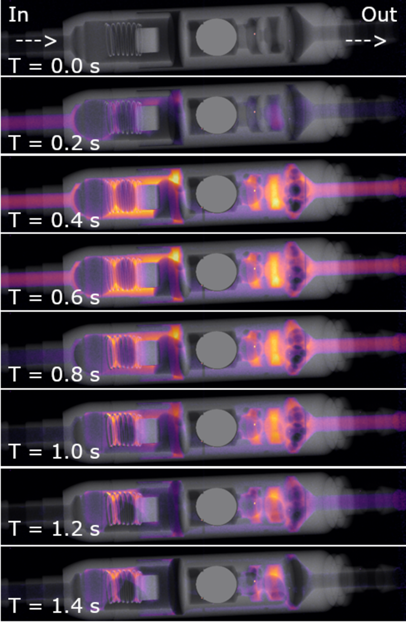
Time-resolved imaging of contrast agent solution flow through a cerebro-spinal fluid shunt valve.