Super-sampled micro-radiography
Photon-counting detectors
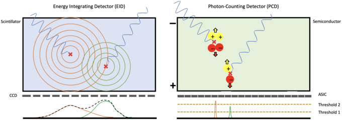
As opposed to state-of-the-art scintillating detectors, photon-counting detectors directly convert excitons (charge carrier pairs generated by ionizing radiation) into detector signal. This is achieved by separating the charge carriers by charge sign through an external electrical field. Fast electronics within each pixel are consecutively used to analyze signals generated by individual X-ray interactions, giving spectral and timing information.
Since the motion of the secondary signal carriers, i.e. the generated charge carriers is directed by an external field, the lateral spread within the sensor is small. In contrast, secondary photons generated in a scintillator propagate isotropically within the sensor, leading to significant lateral smearing and a detrimental trade-off between sensor thickness, and therefore absorption efficiency, and image contrast.
Super-resolution imaging
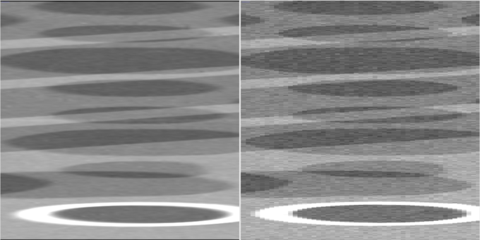
The inherently high contrast of photon-counting detectors allows for efficient super-resolution strategies. An approach to this we implemented at our institution is piezo-driven mechanical super-sampling, followed by iterative deconvolution. To this end any of the elements of the imaging pipeline, i.e. the X-ray source, sample or detector can be shifted by miniscule sub-pixel steps. Since construction of mechanically reliable motion systems at this precision is challenging, we instead implemented a software-driven shift-detection with sub-pixel precision.
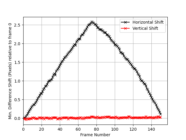
Consecutive frames taken within the super-sampled series are co-registered to a key frame prior to image deconvolution. The above figure illustrates the detected shift in case a ramp signal is applied to one of the XY-motion systems.
As a result of this imaging strategy, we obtain about 10x higher feature resolution for detectors of the EIGER2X-1M CdTe and Santis-500k GaAs detectors.
Dr. Martin Peter Pichotka
Scientific Lead of the Spectral CT Research Group
Tel.: +49 761 270-39220
E-Mail: martin.pichotka@uniklinik-freiburg.de
Medical Center – University of Freiburg
Dept. of Radiology · Medical Physics
Killianstr. 5a
79106 Freiburg


