CT imaging
CT is a 3D imaging modality, based on the acquisition of a series of radiographs and their consecutive volumetric reconstruction. The result of a classic CT scan yields a volumetric representation of the patient / sample featuring grey value encoded CT numbers, which in turn represent the local material density multiplied by the convolution of the X-ray spectrum employed and the respective material’s energy dependent X-ray attenuation coefficient. Given a sufficiently homogeneous material composition and narrow X-ray spectrum, the respective gray values give an approximate measure of local material density.
Key characteristics in classic CT imaging are:
- Achievable volumetric resolution
- Contrast between constituent tissue types / materials
- Required patient dose / machine time
- Noise level within the volumetric image
- Presence and magnitude of imaging artefacts
These characteristics are influenced by a number of factors, including:
- Detector characteristics (stopping power, contrast, MTF, energy resolution in case of spectral detectors, dark current, …)
- Volumetric reconstruction algorithms. Here two main classes exist:
Many of todays CT machines still employ direct inversion based aglortihms (FBP, FDK, …), which are computationally inexpensive and fairly easy to integrate.
Iterative reconstruction techniques are computationally more expensive, which, in particular
Our group focuses on energy resolved CT imaging, exploiting the superior characteristics of the emerging photon counting detector technology.
As opposed to current state-of-the-art scintillator based detector technology, with photon counting technology single ionizing interactions with the sensor are processed individually, permitting determination of photon energy and time-of-arrival in radiographic applications. This in principle allows to not only determine local attenuation factors, represented as gray-level values in standard CT, but to extract spectroscopic absorption fingerprints of constituent materials. As a result, different materials and their respective densities can be identified in spectral CT-imaging independently.
Furthermore, spectral CT imaging inherently permits to suppress beam hardening artefacts, which in presence of high attenuation structures (e.g. metallic implants) in standard CT imaging have severe detrimental effects on the image quality. Simultaneously, the soft tissue contrast, which in classic CT applications is low due to X-ray spectrum truncation towards low photon energies, can be enhanced by utilization of broad spectra.
In combination with (targeted) contrast agents, quantitative material decomposition in spectral CT permits to perform functional imaging, therefore dramatically extending the range of potential CT applications in bio-medical imaging.
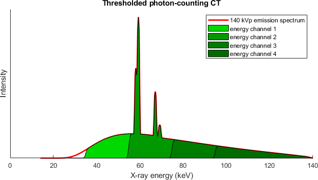
Energy Binning vs. Beam-Filtering
Since broad X-ray spectra, without spectral data acquisition and processing, lead to severe beam hardening artefacts, a straightforward solution of the problem is to narrow down the spectra employed in CT imaging. Synchrotron light sources and free electron lasers represent this concept its most extreme manifestation. These huge, multi-billion Euro facilities are capable of producing quasi-monochromatic X-ray beams, but also of extreme brightness and with small angular deviation (denoted as brilliance). However, such sources are completely impractical for medical and laboratory use.
In clinical CT typically high powered X-ray tubes (several to hundreds to kW) are employed in combination with heavy beam filtering (Al, Cu, Sn). Although providing the desired mono-chromatization of the tube spectrum, the filtering entails several adverse effects. For one thing the loss in brightness entails a trade-off between spectrum width, exposure time (hence motion artefacts) and required spot size (hence system resolution). For the other, with the spectra used in clinical setting, beam-hardening artefacts from bone structures are largely suppressed, however at the cost of soft tissue contrast.
Filtered (red) vs. energy-binned (shaded green) X-ray spectrum. Other than permitting discrimination of X-ray quanta by energy, the application of photon counting detectors in CT allows to utilize a larger portion of the X-ray tubes photon flux.
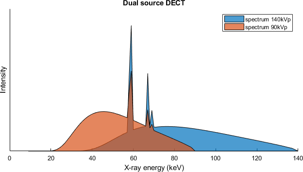
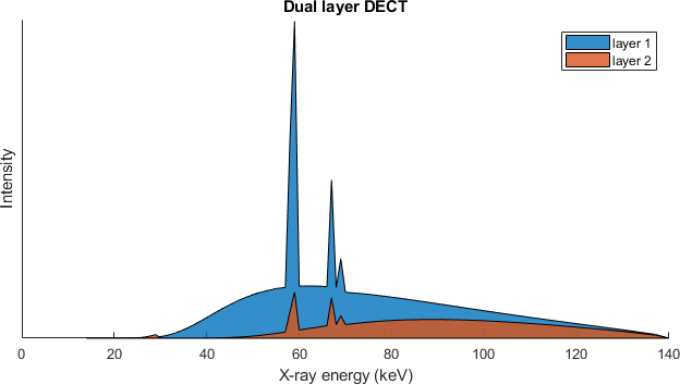
Spectral CT vs. Dual-Energy CT
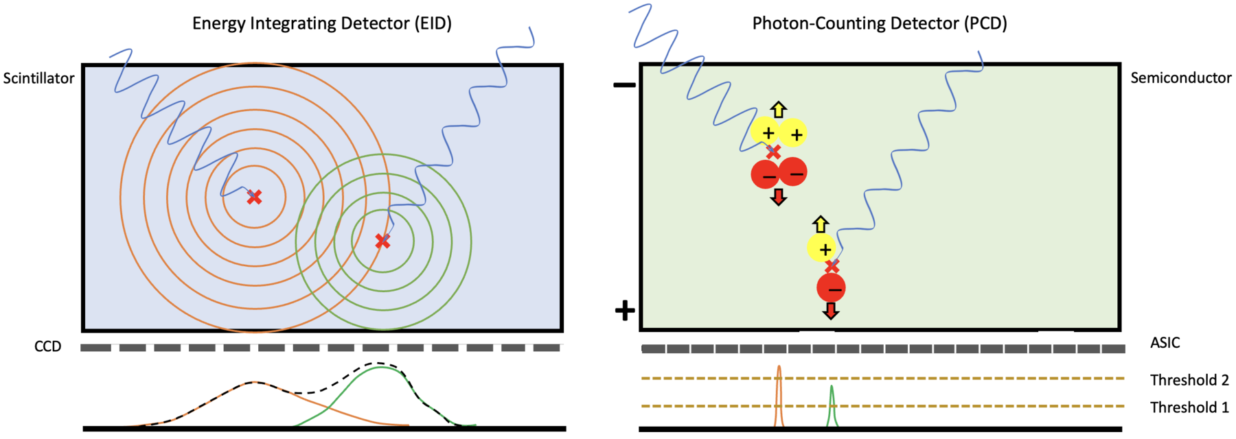
Photon-counting detectors
As opposed to state-of-the-art scintillating detectors, photon-counting detectors directly convert excitons (charge carrier pairs generated by ionizing radiation) into detector signal. This is achieved by separating the charge carriers by charge sign through an external electrical field. Fast electronics within each pixel are consecutively used to analyze signals generated by individual X-ray interactions, giving spectral and timing information.
Since the motion of the secondary signal carriers, i.e. the generated charge carriers is directed by an external field, the lateral spread within the sensor is small. In contrast, secondary photons generated in a scintillator propagate isotropically within the sensor, leading to significant lateral smearing and a detrimental trade-off between sensor thickness, and therefore absorption efficiency, and image contrast.

Dr. Martin Peter Pichotka
Scientific Lead of the Spectral CT Research Group
Tel.: +49 761 270-39220
E-Mail: martin.pichotka@uniklinik-freiburg.de
Medical Center – University of Freiburg
Dept. of Radiology · Medical Physics
Killianstr. 5a
79106 Freiburg