Research group Tebartz van Elst / Nickel
Visual Neuroscience| Research group leader: | Prof. Dr. Ludger Tebartz van Elst Phone: +49 (0)761 270 - 65010 Email: tebartzvanelst@uniklinik-freiburg.de Prof. Dr. Kathrin Nickel Phone: +49 (0)761 270 – 65010 Email: kathrin.nickel@uniklinik-freiburg.de Secretariat: Frau Griesbaum Phone: +49 (0)761 270 - 65290 |
|---|---|
| Project coordinator: | Evelyn Friedel, M.Sc. Phone: +49 (0)761 270 – 69081 Email: evelyn.friedel@uniklinik-freiburg.de |
| Group members: | Prof. Dr. Dieter Ebert (MD) Prof. Dr. Dominique Endres (MD) Dr. rer. nat. Simon Maier (biologist) Malina Beringer (MD student) Sophie Wiesend (MD student) Katharina Busch (MD student) |
| Cooperation partners: | Prof. Dr. rer. nat. Sven Heinrich (Head of the Section for Functional Vision Research, Eye Center, Medical Center, University of Freiburg) Prof. Dr. Michael Bach (Eye Center, Medical Center, University of Freiburg) Prof. Dr. Wolf A. Lagrèze (Head of the Section Neuroophthalmology, Pediatric Ophthalmology and Strabology, Eye Center, Medical Center, University of Freiburg) Dr. Sebastian Küchlin (MD, Eye Center, Medical Center, University of Freiburg) PD Dr. Emanuel Bubl |
Research focus
The retina and its analysis by routine ophthalmological examination methods have become increasingly important in psychiatric research to investigate potential and valid biomarkers for different psychiatric disorders. As an ontogenetic and easily accessible part of the brain, the retina contains not only specialized neurons but also neurotransmitters of the central nervous system, like dopamine. Alterations in the function of the dopaminergic system are assumed to be linked to different mental disorders, e.g. schizophrenia or depression.
Since retinal signal transmission is modulated by retinal dopaminergic activity, the investigation of early retinal responses might provide information about the integrity and function of the central dopaminergic system.
Investigations of the retinal anatomy, on the other hand, could provide insight into structural alterations of neuronal tissue that is possibly related to neurodegenerative processes, or might indicate neuroinflammation.
We apply two ophthalmological tools for the investigation of the retina:
1. Functional analysis with the electroretinogram (ERG)
The electroretinogram (ERG) is an ophthalmological examination method to measure the electrical activity of different retinal cells, similar to an “ECG” of the heart. Depending on the type of stimulus, the function of different retinal cells can be tested.

2. Structural analysis with the optical coherence tomography (OCT)
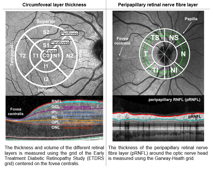
The OCT is an imaging technique similar to ultrasonography, which uses light instead of sound to generate two- and three- dimensional images of the retina. Moreover, OCT allows to measure the different retinal cell layers and their thickness and volume. We focus not only on the area around the fovea centralis, but also on the optic nerve head (papilla) and the thickness of the peripapillary retinal nerve fibre layer (pRNFL).
Current state of our research
In previous studies, we detected that patients suffering from depression show a significant reduction in the PERG signals compared to healthy controls. Based on the alterations on the retinal level, we were able to distinguish depressed patients from healthy controls with high sensitivity and specificity. Furthermore, we could demonstrate that the PERG signal normalized after remission from depression. With regard to the question of objective biomarkers in neuropsychiatry, the processes on the retinal level (measured with the PERG) could represent a valid biomarker for depressive states in humans.
Our recently accomplished studies revealed that patients with schizophrenia have significant alterations in both the fERG and the OCT when compared to healthy individuals. In contrast to this, patients with autism spectrum disorders only showed structural retinal alterations (OCT) and normal retinal functioning (fERG).
Currently, our research group focuses on the investigation of the specificity of this signal in different neuropsychiatric disorders (depression, bipolar disorder, schizophrenia, attention-deficit/hyperactivity disorder (ADHD) and autism spectrum disorder) and in response to various therapeutic interventions (medication, psychotherapy etc.). Moreover, we are currently testing the suitability of a new portable device to measure the flash-ERG which would allow the application in clinical routine. Finally, by comparing both approaches (ERG and OCT), we aim to investigate possible associations between functional and structural retinal alterations.
Research projects
Recently completed:
- Assessment of pattern-ERG in depression
- Normalization of pattern-ERG alterations after remission from depression
- Elevated retinal background noise in ADHD
- Normalization of increased retinal background noise after ADHD treatment
- Normal visual acuity and electrophysiological contrast gain in patients with autism spectrum disorder
- Optimization of the pattern-ERG stimulus parameters for the application in clinical routine
- Functional and structural investigation of the retina in patients with schizophrenia with the fERG and OCT
- Investigation of the correlation of functional and structural retinal alterations in patients with depression applying new approaches e.g. fERG with the RETeval® system and OCT
- Investigation of the application of different electrodes
Current projects:
- Comparison of pattern-ERG and fERG in patients with depression, bipolar disorder and ADHD
- Pharmacological effects of psychiatric medication on the retinal signals and structure
- Investigation of associations between structural retinal alterations and neuroimaging parameters
Planned Projects:
- Identification of the timing when pattern-ERG alterations normalize in the course of the remission from depression
Selected publications
- Nickel K, Tebartz van Elst L, Beringer M, Endres D, Runge K, Maier S, Küchlin S, Bach M, Domschke K, Heinrich SP, Friedel EBN. 2024. Analysis of skin and corneal fiber electrodes for electroretinogram assessments in patients with major depressive disorder. Frontiers in Neuroscience. doi.org/10.3389/fnins.2024.1501149.
- Nickel K, Heinrich SP, Beringer M, Endres D, Runge K, Küchlin S, Maier S, Bach M, Domschke K, Tebartz van Elst L, Friedel EBN. 2024. Alterations in center-surround contrast suppression in patients with major depressive disorder. Scientific Reports. 14(1):28160. doi.org/10.1038/s41598-024-78584-z.
- Friedel EBN, Tebartz van Elst L, Beringer M, Endres D, Runge K, Maier S, Kornmeier J, Bach M, Domschke K, Heinrich SP, Nickel K. 2024. Reduced contrast sensitivity, pattern electroretinogram ratio, and diminished a-wave amplitude in patients with major depressive disorder. European Archives of Psychiatry and Clinical Neuroscience. doi.org/10.1007/s00406-024-01826-8.
- Friedel EBN, Haldina J, Nickel K, Bach M, Tebartz van Elst L, Heinrich SP. Effect of eccentric fixation on the steady-state pattern electroretinogram. 2024. Documenta Ophthalmologica. 148(2):87-95. doi.org/10.1007/s10633-024-09967-w.
- Friedel EBN, Tebartz van Elst L, Schäfer M, Maier S, Runge K, Küchlin S, Reich M, Lagrèze WA, Kornmeier J, Ebert D, Endres D, Domschke K, Nickel K. 2022. Retinal Thinning in Adults with Autism Spectrum Disorder. Journal of Autism and Developmental Disorders. doi.org/10.1007/s10803-022-05882-8
- Friedel EBN, Hahn HT, Maier S, Küchlin S, Reich M, Runge K, Bach M, Heinrich SP, Kornmeier J, Endres D, Ebert D, Domschke K, Tebartz van Elst L, Nickel K. 2022. Structural and functional retinal alterations in patients with paranoid schizophrenia. Translational Psychiatry. 12(1): 402. doi.org/10.1038/s41398-022-02167-7.
- Friedel EBN, Schäfer M, Endres D, Maier S, Runge K, Bach M, Heinrich SP, Ebert D, Domschke K, Tebartz van Elst L, Nickel K. 2022. Electroretinography in adults with high-functioning autism spectrum disorder. Autism Research. 15(11): 2026-2037. doi.org/10.1002/aur.2823.
- Friedel EBN, Tebartz van Elst L, Schmelz C, Ebert D, Maier S, Endres D, Runge K, Domschke K, Bubl E, Kornmeier J, Bach M, Heinrich SP, Nickel K. 2021. Replication of reduced pattern electroretinogram amplitudes in depression with improved recording parameters. Frontiers in Medicine (Lausanne). 8: 732222. doi.org/10.3389/fmed.2021.732222.
- Werner AL, Tebartz van Elst L, Ebert D, Friedel E, Bubl A, Clement HW, Lukačin R, Bach M, Bubl E. 2020. Normalization of increased retinal background noise after ADHD treatment: A neuronal correlate. Schizophrenia Research. 219: 77 – 83. doi.org/10.1016/j.schres.2019.04.013.
- Bubl E, Dörr M, Riedel A, Ebert D, Philipsen A, Bach M, Tebartz van Elst L. 2015. Elevated Background Noise in Adult Attention Deficit Hyperactivity Disorder Is Associated with Inattention. PLoS One 10 (2): e0118271. doi.org/10.1371/journal.pone.0118271.
- Tebartz van Elst L, Bach M, Blessing J, Riedel A, Bubl E. 2015. Normal Visual Acuity and Electrophysiological Contrast Gain in Adults with High-Functioning Autism Spectrum Disorder. Frontiers in Human Neuroscience. 9: 460. doi.org/10.3389/fnhum.2015.00460.
- Bubl E, Dörr M, Philipsen A, Ebert D, Bach M, Tebartz van Elst L. 2013. Retinal Contrast Transfer Functions in Adults with and without ADHD. Edited by Mark W. Greenlee. PLoS One 8 (5): e61728. doi.org/10.1371/journal.pone.0061728.
- Bubl E, Ebert D, Kern E, Tebartz van Elst L, Bach M. 2012. Effect of Antidepressive Therapy on Retinal Contrast Processing in Depressive Disorder. The British Journal of Psychiatry 201 (2): 151–58. doi.org/10.1192/bjp.bp.111.100560.
- Bubl E, Kern E, Ebert D, Bach M, Tebartz van Elst L. 2010. Seeing Gray When Feeling Blue? Depression Can Be Measured in the Eye of the Diseased. Biological Psychiatry. 68 (2): 205–8. doi.org/10.1016/j.biopsych.2010.02.009.
- Bubl E, Tebartz van Elst L, Gondan M, Ebert D, Greenlee MW. 2009. Vision in Depressive Disorder. The World Journal of Biological Psychiatry. 10 (4 Pt 2): 377–84. doi.org/10.1080/15622970701513756.