Tissue Phase Mapping
Marius Menza
We are developing MR-methods based on Phase Contrast Velocity Mapping techniques (Tissue Phase Mapping, TPM) that allow for the acquisition and investigation of myocardial motion with high spatial and temporal resolution. A black blood k-space segmented gradient echo sequence with first-order flow compensation was used for data acquisition. Velocity encoding was performed by adding a bipolar gradient with a venc of 15 cm/s in-plane and 25 cm/s through-plane according to typical velocity values occuring in the wall motion of the left ventricle. In order to overcome limitations in temporal resolution, advanced navigator gating was performed using two navigator echoes per cardiac cycle in combination with real-time acceptance criteria based on signal from successive navigator echo pairs, as recently described [1].
Data post-processing was performed using customized software programmed in Matlab (The Mathworks Inc., Natick, MA). Following contour segmentation and bulk motion correction, the measured in-plane velocities were transformed into an internal polar coordinate system positioned at the center of mass of the segmented left ventricle. As a result, motional parameters are decribed in terms of a cylindrical coordinate system based on radial (contraction / expansion), circumferential (rotation) and longitudinal (shortening / lengthening) velocities leading to a more adapted representation of myocardial motion.
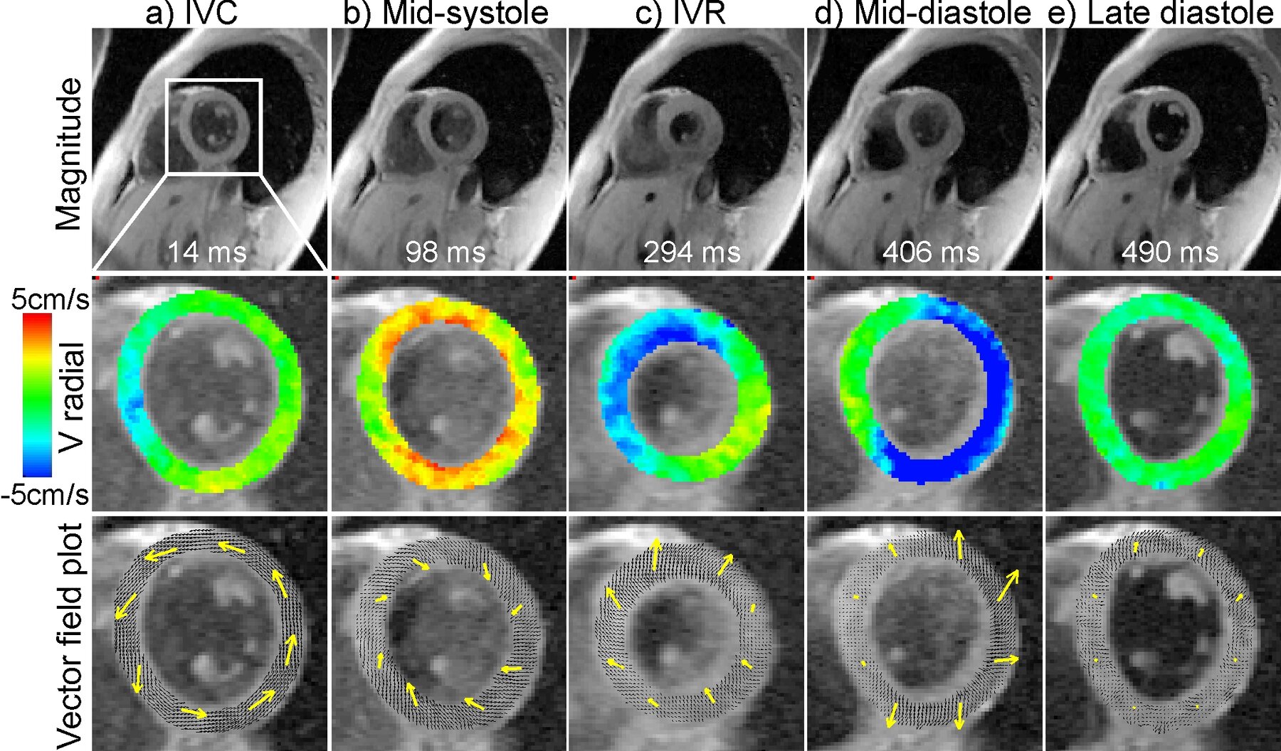
The figure below shows magnitude images (upper row) in a basal slice of a data set with a temporal resolution of 13.8 ms acquired during free-breathing. Five characteristical cardiac frames are shown: early systole during (a,IVC), mid-systole (b), early diastole (c,IVR), mid-diastole (d) and late diastole (e). Corresponding color-coded maps of radial velocities (red: positive velocities, contraction) are shown in the middle row and pixelwise arrow plots of in-plane velocity vector fields in the lower row demonstrating the characteristical in-plane motion of the left ventricle [1].
Analysis of Myocardial Motion: Postprocessing Strategies
The spatial arrangement of myocardial fiber structure affects the mechanical and electrical properties of the heart. Therefore, information on the structure and dynamics of the orientation of the muscle fibers in the human heart might provide significant insight into principles of the mechanics of ventricular contraction and electrical propagation and may aid pre- and postsurgical evaluation of patients. Fiber orientation is inherently linked to cardiac wall motion, which can be measured with phase contrast MRI (Tissue Phase Mapping, TPM). Here, this technique was used to generate acceleration fields in order to derive surrogate parameters describing the fiber structure of the left ventricle. High temporal resolution TPM measurements were performed during free-breathing. Based on the assumption that the accelerations are related to the forces generated by the muscle fibers a tracking algorithm was applied to acceleration fields derived from the velocity data for different cardiac frames of left ventricular performance [2].
References
[1] B. Jung, M. Zaitsev, J. Hennig, M. Markl. Navigator Gated High Temporal Resolution Tissue Phase Mapping of Myocardial Motion. Magn Reson Med 2006; 55:937-942.
[2] B. Jung, B. Kreher, M. Markl, J. Hennig. Visualization of Tissue Velocity Data from Cardiac Wall Motion Measurements with Myocardial Fiber Tracking: Principles and Implications for Cardiac Fiber Structures. Eur J Cardiothorac Surg 2006, EPub.

Prof. Dr. Dr. h.c. Jürgen Hennig
Tel.: +49 761 270-74120
University Medical Center Freiburg
Dept. of Radiology · Medical Physics
Killianstr. 5a
79106 Freiburg