
More than "just" a headache
The impact of spinal CSF leaks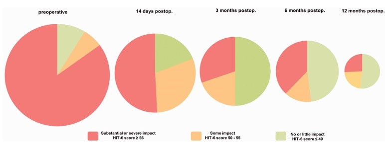
Volz et al. Cephalalgia 2023
SIH and PDPH are widely unknown, under-, and misdiagnosed. „It‘s just a headache“ – that is a common perception. Many patients spent months, even years of their lifes before the diagnosis of this treatable disease is made.
AIMs
- to assess the entire spectrum of the syndrome
- to assess health burden in spinal CSF leaks
to asess outcome that is relevant to the patient
SEAL - Prospective Registry on CSF leaks (#NCT06711731)
Contact & Collaboration
If you are a researcher, or institution interested in joining our initiative, or a patient seeking information, please contact our research team at:
📧 nez.SEAL-registry@uniklinik-freiburg.de
Publications:
Volz et al. Toward quantifying disability in spontaneous intracranial hypotension: A patient-derived disability weight. Cephalalgia, 27April 2026m https://doi.org/10.1177/03331024261444662
Omer et al. Return to work – estimated socioeconomic impact of spontaneous intracranial hypotension and effects of neurosurgical treatment. Front. Neurol., 29 January 2026 https://doi.org/10.3389/fneur.2026.1738826
Wolf K, et al. Mild cognitive impairment in spontaneous intracranial hypotension and its rapid reversal by repair of a spinal cerebrospinal fluid leak. Headache. 2024 Dec 15. doi: 10.1111/head.14882 Epub ahead of print. PMID: 39676275.
Urbach, H., et al. Reply: Dementia in spontaneous intracranial hypotension: look at the spine. Neuroradiology (2024). https://doi.org/10.1007/s00234-024-03529-2
Kraus LM, et al. Chronic post-dural puncture headache-a serious and underrated complication following lumbar puncture: a cohort study. Front Neurol. 2024 Nov 29;15:1493303. doi: 10.3389/fneur.2024.1493303. PMID: 39677860; PMCID: PMC11638535
Volz et al. More than a headache—somatic and mental symptom burden in spontaneous intracranial hypotension before and after surgical treatment. Front. Neurol., 08 October 2024. doi.org/10.3389/fneur.2024.1421579
Urbach et al. Spinal dementia: Don't miss it, it's treatable. Neuroradiology. 2024 July 10. doi:10.1007/s00234-024-03425-9
Volz F, et al. Impact of Spinal CSF Leaks on Quality of Life and Mental Health and Long-Term Reversal by Surgical Closure. Neurol Clin Pract. 2024 Apr;14(2):e200272. doi: 10.1212/CPJ.0000000000200272
Volz, F. et al. Don’t delay, but don’t despair: symptom duration, comorbidity and outcome after closure of spinal cerebrospinal fluid leaks. J Neurol (2024). https://doi.org/10.1007/s00415-024-12242-2
Volz F, et al. Recovery and long-term outcome after neurosurgical closure of spinal CSF leaks in patients with spontaneous intracranial hypotension. Cephalalgia. 2023 Aug;43(8):3331024231196808. doi: 10.1177/03331024231195830.
Jesse et al. Patient-reported symptomatology and its course in spontaneous intracranial hypotension - Beware of a chameleon. Clin Neurol Neurosurg. 2023 Dec 19:236:108087.doi: 10.1016/j.clineuro.2023.108087
Kraus LM et al. Chronic post-dural puncture headache – a serious and underrated complication with a major impact on social life. Abstract. Brain and Spine 2923 3:101958. doi: 10.1016/j.bas.2023.101958
Dobrocky T, et al.. Spontaneous intracranial hypotension: searching for the CSF leak. Lancet Neurol. 2022 Apr;21(4):369-380. doi: 10.1016/S1474-4422(21)00423-3.
Jesse CM, et al. The impact of spontaneous intracranial hypotension on social life and health-related quality of life. J Neurol. 2022 Oct;269(10):5466-5473. doi: 10.1007/s00415-022-11207-7.
Review:
Zander C. et al. Spontaneous intracranial hypotension - a spinal disease. RöFo 2024 June, doi:10.1055/a-2318-8994
Doria-Medina R, Volz F, Lützen N, Beck J. ABC om Spontan intrakraniell hypotension [Spontaneous intracranial hypotension]. Lakartidningen. 2023 Oct 2;120:23092. Swedish. PMID: 37782313.
Urbach H,et al. Spontaneous Intracranial Hypotension. Dtsch Arztebl Int. 2020 Jul 6;117(27-28):480-487. doi: 10.3238/arztebl.2020.0480.
Therapy
What can we do better?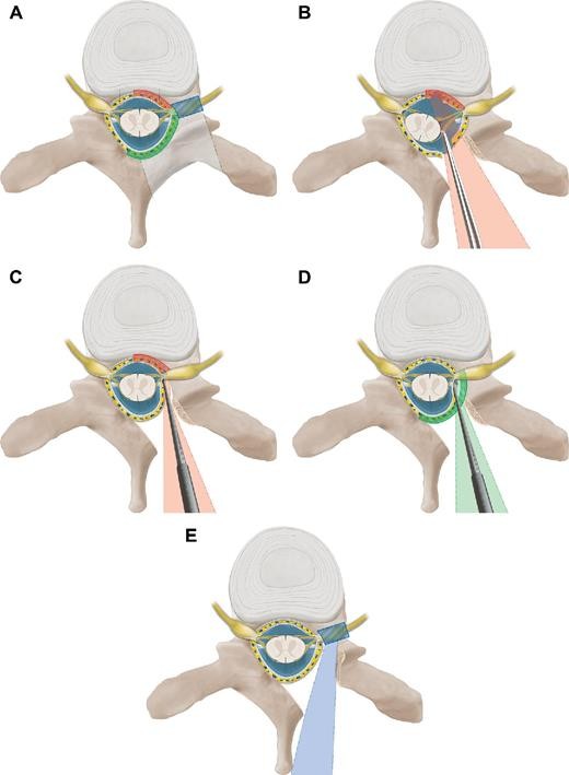
Beck et al. 2018
Spinal CSF Leaks
Spinal CSF leaks can and should be treated. First attempt for symptomatic releave should be a high-volume bloodpatch. In PDPH this can essentially heal the patient.
In case of persistent CSF leak, as in SIH, we aim for a targeted approach to prevent long-term sequelae as subdural hematoma, superficial siderosis, bibrachial amyotrophy, and coma.
AIMs
- to sufficiently seal the leak at lowest risks
Publications:
Overstijns et al. Management of chronic subdural hematoma in spontaneous intracranial hypotension. Brain & Spine, 2025, https://doi.org/10.1016/j.bas.2025.104320
Vasilikos et al. Autologous platelet-rich fibrin as an alternative epidural patch for persistent post-dural puncture headache: A single-center observational study. Interven. Neurorad. 2025, https://doi.org/10.1177/159101992513395
El Rahal et al: Safety, Sequelae, and Efficacy of Nerve Root Clipping in Patients With Spontaneous Spinal Cerebrospinal Fluid Leaks. Operative Neurosurgery ():10.1227/ons.0000000000001401, October 22, 2024. | DOI: 10.1227/ons.0000000000001401
Volz et al. Keyhole Fenestration for Cerebrospinal Fluid Leaks in the Thoracic Spine: Quantification of Bone Removal and Microsurgical Anatomy. Operative Neurosurgery. December 22, 2023. doi: 10.1227/ons.0000000000001042
Piechowiak EI, et al. Epidural Blood Patching in Spontaneous Intracranial Hypotension-Do we Really Seal the Leak? Clin Neuroradiol. 2023 Mar;33(1):211-218. doi: 10.1007/s00062-022-01205-7.
Beck J, et al. Minimally invasive surgery for spinal cerebrospinal fluid leaks in spontaneous intracranial hypotension. J Neurosurg Spine. 2022 Sep 9;38(1):147-152. doi: 10.3171/2022.7.SPINE2252.
Häni L, et al. Outcome after surgical treatment of cerebrospinal fluid leaks in spontaneous intracranial hypotension-a matter of time. J Neurol. 2022 Mar;269(3):1439-1446. doi: 10.1007/s00415-021-10710-7.
Beck J, et al. Posterior Approach and Spinal Cord Release for 360° Repair of Dural Defects in Spontaneous Intracranial Hypotension. Neurosurgery. 2019 Jun 1;84(6):E345-E351. doi: 10.1093/neuros/nyy312. PMID: 30053151.
Beck J, et al. Diskogenic microspurs as a major cause of intractable spontaneous intracranial hypotension. Neurology. 2016 Sep 20;87(12):1220-6. doi: 10.1212/WNL.0000000000003122.
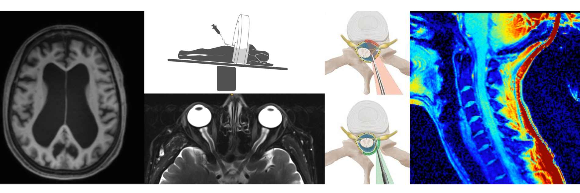
Imaging
Expand the perspectiveOur imaging-based approaches are founded on successful collaboration of several departments:
In the Department of Neuroradiology, the team of Prof. Dr. Urbach and Dr. Lützen work continously to improve different techniques to detect spinal CSF leaks, With the Department of Nuclear Medicine and Prof. Dr. Dr. Meyer, we aim to improve diagnostic marker for spinal CSF leaks.
In collaboration with the Devision of Medical Physics, Prof. Dr. Zeitsev, Dr. Reisert, Department of Neurosurgery, Dr. Wolf, Department of Neuroradiology, Prof. Urbach, Dr. Demerath - automated analysis and postprocessing pipelines (www.nora-imaging.org), non-invasive dynamic measures (phase-contrast MRI), and diffusion microstructure imaging (DMI) are applied to extend our knowledge on pathomechanisms and to explore further imaging-derived biomarker.
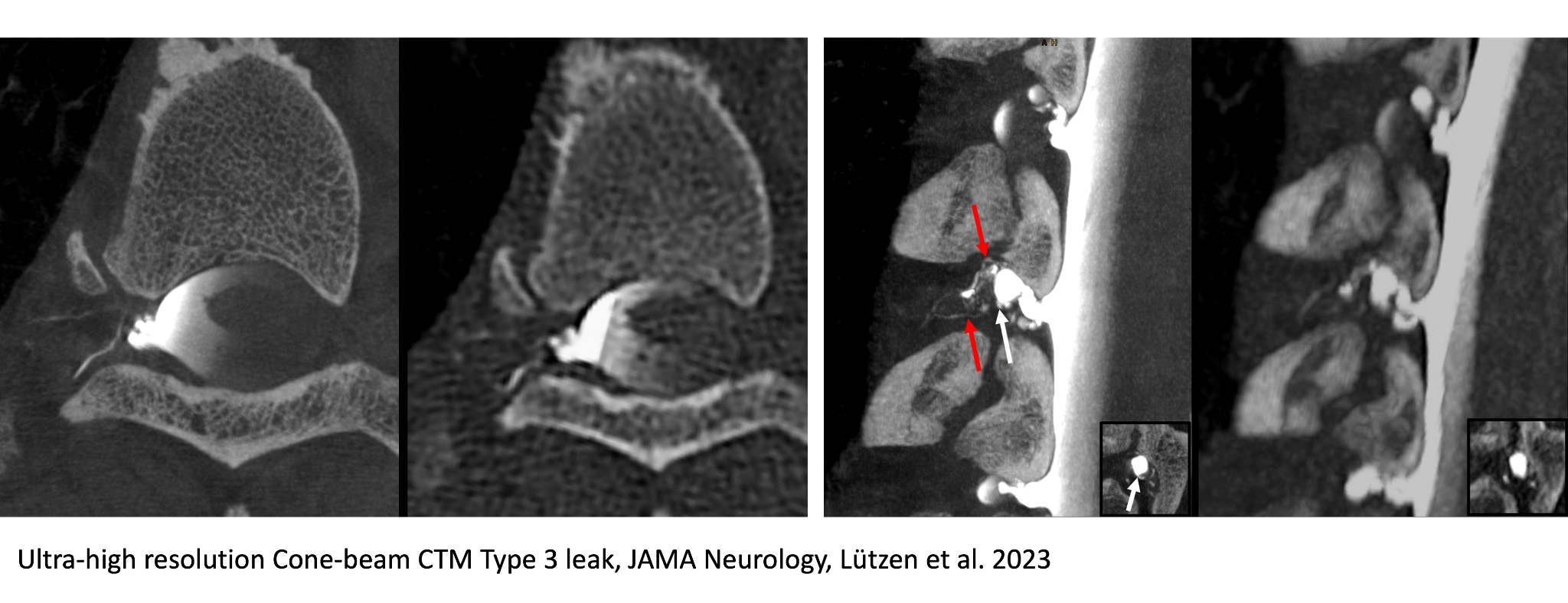
Detection of spinal CSF leaks
It is our goal to investigate new methods, and ultimately improve sensitivity and specificity to detect spinal CSF leaks. While SIH can be detected in most cases, we are still nearly „blind“ detecting PDPH.
AIMs
- to better understand pathomechanisms
- to reduce radiation dose
- to improve diagnostic pathways
- to improve reliability
Publications:
Zander et al. Volumetric response after closure of a spinal CSF leak in patients with spontaneous intracranial hypotension: a multicompartmental longitudinal study. Journal of NeuroInterventional Surgery 2026;18:241-247.
Lützen et al. Temporal Characteristics of Type 2 Lateral Spinal CSF Leaks on Digital Subtraction Myelography: Fast, Medium or Slow Leaks? AJNR Oct 2025, ajnr.A9040; DOI: 10.3174/ajnr.A9040
Zander et al. Patients with chronic post-dural puncture headache do not have typical imaging features of intracranial hypotension: An MRI study using the Bern score. Headache 2025 https://doi.org/10.1111/head.15057
Lützen et al. Advanced Imaging of Type 2 Spinal CSF Leaks with Ultra-High-Resolution Conebeam CT Myelography. AJNR 2025, Jun 2025, DOI: 10.3174/ajnr.A8675
Lützen et al. Digital Subtraction Myelography for the Detection of Type 1 Spinal CSF Leaks: Evaluation of Temporal Characteristics and Diagnostic Value. AJNR 2025, ajnr.A8847; DOI: https://doi.org/10.3174/ajnr.A8847
Lützen et al. MRI and Surgical Findings Refine Concepts of Type 2 Cerebrospinal Fluid Leaks in Spontaneous Intracranial Hypotension. Radiology Feb. 2025 https://doi.org/10.1148/radiol.241653
Lützen et al. Advanced Imaging of Type 2 Spinal CSF Leaks with Ultrahigh-Resolution Cone-Beam CT Myelography. AJNR 2025, ajnr.A8675; DOI: https://doi.org/10.3174/ajnr.A8675
Madhavan AA, Lutzen N, et al. Additional Diagnostic Value of Cone Beam CT Myelography Performed After Digital Subtraction Myelography for Detecting CSF-venous Fistulas. AJNR Am J Neuroradiol. 2024 Oct 15:ajnr.A8535. doi: 10.3174/ajnr.A8535 Epub ahead of print. PMID: 39406513.
Lützen et al. Primary CSF-lymphatic fistula: a previously unknown cause of spontaneous intracranial hypotension. J Neurol 271, 7016–7020 (2024). https://doi.org/10.1007/s00415-024-12598-5.
Lützen N, Demerath T, Volz F, Beck J, Urbach H. Cone-Beam CT for the Detection of a Ventral Spinal CSF Leak in Spontaneous Intracranial Hypotension. Neurology. 2023 Jul 27:10.1212/WNL.0000000000207504.
Lützen N, et al. Prone Dynamic CT Myelography in Spontaneous Intracranial Hypotension: Diagnostic Need and Radiation Doses. Clin Neuroradiol. 2023 Sep;33(3):739-745. doi: 10.1007/s00062-023-01269-z.
Lützen N, Beck J, Urbach H. Cerebrospinal Fluid Venous Fistula Imaging with Ultrahigh-Resolution Cone-Beam Computed Tomography. JAMA Neurol. 2023 Aug 1;80(8):870-871.
Lützen N, et al. Sacral Dural Tears as a Cause of Spontaneous Intracranial Hypotension. Clin Neuroradiol. 2023 Jun 1. doi: 10.1007/s00062-023-01292-0.
Lützen N, et al.. Spontaneous intracranial hypotension: diagnostic and therapeutic workup. Neuroradiology. 2021 Nov;63(11):1765-1772. doi: 10.1007/s00234-021-02766-z
Arnold PG, et al. Support Vector Machine-based Spontaneous Intracranial Hypotension Detection on Brain MRI. Clin Neuroradiol. 2022 Mar;32(1):225-230.
Evangelou P, et al. 68Ga-DOTA PET for Diagnosis of Spinal Cerebrospinal Fluid Leaks. J Nucl Med. 2023 Mar;64(3):430-436. doi: 10.2967/jnumed.122.264059
Biomarker
machanics or metabolism?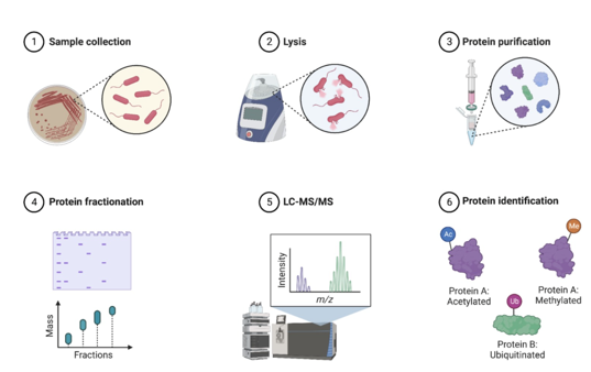
Why does a person develop SIH or PDPH? Are there risk factors? Do we need to look beyond mechanics? Might we be able to detect and monitor spinal CSF leaks by a drop of blood?
The analysis of fluid-derived biomarker, and tissue will help to answer these questions.
AIMs
- to monitor markers for superficial siderosis in spinal CSF leaks, which is a common long-time sequelae of intracranial hypotension causing neurological deterioration.
- to implement proteomics and transcriptomics to depict possible diagnostic and therapeutic targets
Current Project:
The objective of the P-InSIHght study is to explore the utility of proteomics as a diagnostic and investigative tool in SIH. Through large-scale protein analysis of samples from SIH patients and controls, the research group aim to identify biomarkers that can provide insight into the pathology of SIH and potentially serve as a non-invasive diagnostic criterion and even as a follow-up tool.
Publications:
Häni L, et al. Cerebrospinal fluid biomarkers of superficial siderosis in patients with spontaneous intracranial hypotension. Eur J Neurol. 2023 Jan;30(1):235-240. doi: 10.1111/ene.15591. Epub 2022 Oct 19. PMID: 36209476.
El Rahal A, et al. Surgical closure of spinal CSF leaks improves symptoms in patients with superficial siderosis. Eur J Neurol. 2023 Nov 28. doi: 10.1111/ene.16122.
Histology
tackling pathomechanisms in spinal CSF leaks
Understanding spinal CSF leaks
In collarboration with the Institute for Neuropathology, we aim to understand the pathomechanisms of spinal CSF leaks. What happens inside the spinal canal that prevents the dura from healing? what is the exact histology of CSF leaks?
AIMs
- to better understand pathomechanisms of spinal CSF leaks
- to improve diagnostic pathways
- to improve treatment pathways
Publications:
Häni L et al. Distinct Pattern of Membrane Formation With Spinal Cerebrospinal Fluid Leaks in Spontaneous Intracranial Hypotension. Operative Neurosugery 2023. Sep doi: 10.1227/ons.0000000000000914
CSF Circulation
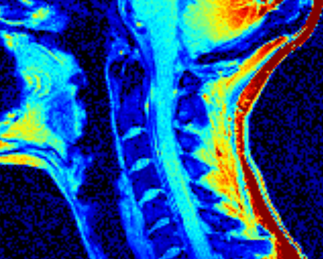
phase-contrast MRI - color-coded velocities in head-feed direction
The traditional concepts of CSF production, resorption, and function have been challenged over the last years. The maintainance of CSF circulation is beliefed as being crucial for the function of the brain. Thus, understanding these machnisms is highly important in diseases related to impaired CSF circulation.
There are few non-invise approaches, as phase-contrast MRI, that allow a glimps into the system without any outer manipulation.The standard procedure to investigate parameters of CSF circulation is infusion testing.
In disorders with impaired CSF circulation, the infusion test serves as a diagnostic tool based on established markers. Yet many aspects are not fully understood.
AIMs
- to investigate CSF circulation of the craniospinal system
- to investigating non-invasive tools as potential substitutes
Publications:
Fung et al. CSF Pressure and Dynamics in Patients With Chronic Postdural Puncture Headache
A Single-Center Cohort Study. Neurology, 2025. https://doi.org/10.1212/WNL.0000000000213998
Urbach H, et al. Different Glymphatic Kinetics in Spontaneous Intracranial Hypotension. AJNR Am J Neuroradiol. 2024 Oct 3;45(10):1605-1612. doi: 10.3174/ajnr.A8365.
Wolf K, et al. Non-invasive biomarkers for spontaneous intracranial hypotension (SIH) through phase-contrast MRI. J Neurol (2024). https://doi.org/10.1007/s00415-024-12365-6
Wolf K, et al. CSF Flow and Spinal Cord Motion in Patients With Spontaneous Intracranial Hypotension: A Phase Contrast MRI Study. Neurology. 2023 Feb 14;100(7):e651-e660. doi: 10.1212/WNL.0000000000201527.
Beltrán S, et al. Spinal cord motion and CSF flow in the cervical spine of 70 healthy participants. NMR Biomed. 2023 Aug 3:e5013. doi: 10.1002/nbm.5013.
Wolf K, et al. Idiopathische intrakranielle Hypertension und Phasenkontrast-MRT: ein neuer Ansatz. Clin Neuroradiol 33 (Suppl 1),1-140(2023).https://doi.org/10.1007/s00062-023-01336-5
Wolf K, et al. Spinal cord motion assessed by phase-contrast MRI - An inter-center pooled data analysis. Neuroimage Clin. 2023;37:103334. doi: 10.1016/j.nicl.2023.103334.
Karimi F, et al. Theory for a non-invasive diagnostic biomarker for craniospinal diseases. Neuroimage Clin. 2023;37:103280. doi: 10.1016/j.nicl.2022.103280.
Kraus LM et al. A chronic post dural puncture syndrome without low opening pressure or altered CSF dynamics. Abstract. Brain and Spine 3:101914. doi:10.1016/j.bas.2023.101914
Wolf K, et al. Focal cervical spinal stenosis causes mechanical strain on the entire cervical spinal cord tissue - A prospective controlled, matched-pair analysis based on phase-contrast MRI. Neuroimage Clin. 2021;30:102580. doi: 10.1016/j.nicl.2021.102580
Wolf K, et al. Spinal Cord Motion in Degenerative Cervical Myelopathy: The Level of the Stenotic Segment and Gender Cause Altered Pathodynamics. J Clin Med. 2021 Aug 25;10(17):3788. doi: 10.3390/jcm10173788.
Häni L, et al. Insights into the natural history of spontaneous intracranial hypotension from infusion testing. Neurology. 2020 Jul 21;95(3):e247-e255. doi: 10.1212/WNL.0000000000009812.
Beck J, et al. Cerebrospinal fluid outflow resistance as a diagnostic marker of spontaneous cerebrospinal fluid leakage. J Neurosurg Spine. 2017 Aug;27(2):227-234. doi: 10.3171/2017.1.SPINE16548.
Research requires strong collaborations across specialties and borders. This page represents efforts of the interdisciplinary CSF Team in collaboration with
University of Freiburg:
Department of Neurosurgery
Department of Neuroradiology
Department of Radiology, Medical Physics
Department of Nuclear Medicine
Department of Neuropathology
Department of Anaesthesiology
Department of Opthalmology, Section Neuroopthalmology
Swiss:
Department of Neuroradiology, Inselspital, Bern
Department of Neurosurgery, Inselspital Bern
The interface group, UZH Zürich
UK:
King's College, London
University College of London
USA:
Stanford Medical, CA
Cedars-Sinai Medical Center, CA
Denmark:
Danish Headache Center, Copenhagen
Klinik für Neurochirurgie
im Neurozentrum
Breisacher Straße 64
D-79106 Freiburg
Telefon: 0761 270-50010 /-50020
Telefax: 0761 270-50240
neurochirurgie@uniklinik-freiburg.de

Prof. Dr. Jürgen Beck
Ärztlicher Direktor
Telefon: 0761 270-50060
International call: +49-761-270-50060
j.beck@uniklinik-freiburg.de

Marc Mielke
Pflegedienstleiter
Tel. +49 761 270 52260
FAX +49 761 270 54200
marc.mielke@uniklinik-freiburg.de

Christian Böke
Kaufmännische Geschäftsführung Neurozentrum
Telefon: +49 761 270-21071
christian.boeke@uniklinik-freiburg.de