Postprocessing

Detection of White Matter Lesions in the Brain
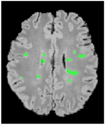
MRI of cerebral white matter may show regions of signal abnormalities. These changes may be associated with hypertension, inflammation, or ischemia, as well as altered brain function. These abnormalities are attributed to lesions and the goal of this work has been to identify them. The data was from elderly normal control subjects or with mild cognitive impairment, and subjects with cerebrovascular disease. The analysis uses 4T FLAIR as well as 4T T1-weighted (T1w) brain images. The pre-processing steps include intensity correction, intra-subject FLAIR to T1w rigid registration. The lesion extraction is performed with region of interest (ROI) identification and subsequent processing of that region for lesion segmentation.
Journal papers and conference proceedings in book form:
- A comparison of different automated methods for the detection of white matter lesions in MRI data;
S. Klöppel, A. Abdulkadir, S. Hadjidemetriou, S. Issleib, L. Frings, T.N. Thanh, I. Mader, S.J. Teipel, M. Hüll, O. Ronneberger
Neuroimage 15, 57(2) 416-22. - Characterization of white matter degeneration in elderly subjects by magnetic resonance diffusion and FLAIR imaging correlation;
Wang Zhan, Yu Zhang, Susanne G. Mueller, Peter Lorenzen, Stathis Hadjidemetriou, Norbert Schuff, Michael W. Weiner,
NeuroImage, February, 2009. - Computational Atlases of Severity of White Matter Lesions in Elderly Subjects with MRI;
Stathis Hadjidemetriou, Peter Lorenzen, Michael Weiner, Susanne Mueller, and Norbert Schuff,
Proc. of 11th International conference on medical image computing and computer assisted intervention, (MICCAI), 2008, September, New York City.
Dr. Marco Reisert
Group Leader
Tel.: +49 761 270-93860
E-Mail: marco.reisert@uniklinik-freiburg.de
University Medical Center Freiburg
Dept. of Radiology · Medical Physics
Killianstr. 5a
79106 Freiburg

