Novel Contrasts
Bayesian Estimation of Diffusion Metrics
Diffusion-sensitized magnetic resonance imaging probes the cellular structure of the human brain, but the primary microstructural information gets lost in averaging over higher-level, mesoscopic tissue organization such as different orientations of neuronal fibers. While such averaging is inevitable due to the limited imaging resolution, we investigate in this project methods for disentangling the microscopic cell properties from the effects of mesoscopic structure based in information principles.
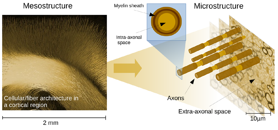
Disentangeling the mesostructural and microstructural components of the dMRI signal is a big challenge. We extract features that dependend only on microstructure to obtain stable white matter diffusion metrics. Computation just takes seconds and applications are manifold and have to explored.
- M Reisert, E Kellner, B Dhital, J Hennig, VG Kiselev
“Disentangling micro from mesostructure by diffusion MRI: a Bayesian approach”
Neuroimage, 2017 - Elsevier, (download the code here) - Elias Kellner, Karl Egger, Valerij G Kiselev, Horst Urbach, and Marco Reisert
“Microstructure Parameters in acute Stroke: A Bayesian approach to diffusion- weighted MRI”
Proceedings of ISMRM 2015, Singapur - Elias Kellner, Marco Reisert, Ori Staszewski, Bibek Dhital , Valerij G Kiselev, Karl Egger , Horst Urbach, and Irina Mader
“Bayesian Estimation of Microstructural Parameters in Glioma Patients, and Comparison with Genetic Analysis”
Proceedings of ISMRM 2015, Singapur - Marco Reisert, Elias Kellner, Bibek Dhital, Juergen Hennig, Valerij G. Kiselev
“Diffusion MRI: Disentangling Micro- from Mesostructure and Bayesian Parameter Evaluation”
Proceedings of ISMRM 2015, Singapur

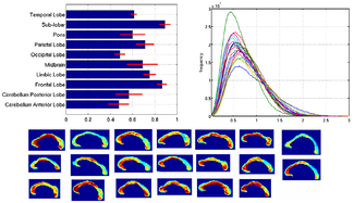
Example maps on a clinical feasible protocol
Tensor divergence
The absolute determination of axonal volume fraction is challenging with respect to measurement techniques and analysis. Unfortunately, the usually employed multi-compartement models cannot be applied to single q-shell measurements, because the compartement’s diffusivities cannot be resolved. However, the relative volume fractions can be inferred up to a certain accuracy. This project proposes an equation for fiber orientation densities that can infer the absolute fraction up to a global factor. This equation, which is inspired by the classical mass preservation law in fluid dynamics, expresses the fiber conservation associated with the assumption that fibers do not terminate in white matter.
The fiber densities derived by tensor divergence show reasonable relative distributions in the corpus callosum. Also in other regions TD derived densities can serve as a reliable biomarker

Comparison of TFD to other contrasts
- Mohammadi, S., Carey, D., Dick, F., Diedrichsen, J., Sereno, M. I., Reisert, M., Weiskopf, N.
“Whole-brain in-vivo measurements of the axonal g-ratio in a group of 37 healthy volunteers.”
Frontiers in neuroscience, 9(2015)
- Marco Reisert, Irina Mader, Roza Umarova, Simon Maier, Ludger Tebartz van Elst and Valerij G. Kiselev
“Fiber Density Estimation from Single Q-Shell Diffusion Imaging by Tensor Divergence”
Neuroimage. 2013 Mar 26;77C:166-176. doi: 10.1016/j.neuroimage.2013.03.032 -
Marco Reisert, Henrik Skibbe and Valerij G. Kiselev
“Tensor Divergence for Fiber Density Estimation”
In Proceedings of MICCAI 2012
Funding source: Deutsche Forschungs Gemeinschaft DFG RE 3286/2-1
Diffusion Textures
The analysis and interpretation of dMRI measurements is a highly ill-posed inverse problem. There is a great number of microstructural models around and conclusive answers are still missing. The challenge of achieving higher specificity by disentangling the dMRI signal into independent tissue properties is not yet mastered. Practical diffusion measurements do not provide enough information for inverting these models and, hence, suffer from instabilities and artifacts. In this project we want to investigate a different viewpoint to the problem. The common approach is reductionistic: a complex biophysical model describing the constituent parts of the tissue and their interactions is set up, and, due to the degeneracy of the dMRI signal, the model is simplified to a practically relevant level. We aim for an alternative representation capturing all properties of the signal in a transparent and comprehensive way. The idea is to turn the originally six-dimensional dMRI signal into a equivalent three-dimensional, high-resolution map putatively representing the microstructure of the tissue. Of course, finding the true microstructurual patch is not possible at all. However, finding one statistical instance of microstructure, which is able to generate the observed dMRI signal is a viable alternative.

Dr. Marco Reisert
Group Leader
Tel.: +49 761 270-93860
E-Mail: marco.reisert@uniklinik-freiburg.de
University Medical Center Freiburg
Dept. of Radiology · Medical Physics
Killianstr. 5a
79106 Freiburg