Investigation of complex blood flow pattern in the mouse aorta.
A new setup was established where a prospectively gated 4D phase contrast sequence is combined with a highly sensitive cryogenic coil on a preclinical magnetic resonance scanner. The sequence was redesigned to maintain a close to steady state condition of the longitudinal magnetization and hence to overcome steady state artifacts. Imaging parameters were optimized to provide high spatial and temporal resolution. Pathline visualizations were generated from the acquired velocity data in order to display complex flow patterns.
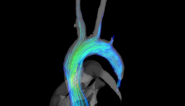
Streamlines in the aorta during systole as described in [1].
Parameters: FOV 20 x 16 x 14 mm³; Matrix 68 x 50 x 50 ; Resolution 160 µm³; TE/TR = 2ms/5 ms; Acquisition time = 40 min
Description and image taken from:
[1] Braig M, Leupold J, Menza M, Russe M, Ko C-W, Hennig J, et al. Preclinical 4D-flow magnetic resonance phase contrast imaging of the murine aortic arch. PLOS ONE. 2017;12: e0187596. doi:10.1371/journal.pone.0187596
Compressed Sensing accelerated radial cardiovascular MRI:
An intravascular gadolinium based contrast agent (100µl+100µl NACL) Gadospin P (nanoPET Pharma GmbH) was applied via tail injection prior to the start of the data acquisition. A preclinical MRI system Bruker BioSpec 70/20 USR equipped with a two channel transmit/receive cryogenic (Bruker) surface coil was used. Respiration (pressure sensor) and ECG were acquired synchronously to the MRI acquisition. A radial 3D center-out sequence was implemented. Parameters: FOV = 22x22x22 mm³, TE/TR = 0.6 ms/3.6 ms; matrix =120x120x120; FA = 8°, Acquisition time in vivo ~14 min. Data were continuously acquired and sorted retrospectively according to the ECG and respiration signals (respiration events are discarded) during reconstruction. A NUFFT- operator based reconstruction in combination with a compressed sensing was used. The microstructure of the heart muscle is clearly visible. Tiny structures that are usually not visible in common cardiovascular MRI can be visualized. The 3D acquisition can be arbitrarily formatted and every part of the heart and the aorta can be visualized as the whole dataset is reconstructed with an isotropic resolution of 90 µm.

apl. Prof. Dr. Dominik von Elverfeldt
Head of AMIR
Tel.: +49 761 270-38320
E-Mail: dominik.elverfeldt@uniklinik-freiburg.de
University Medical Center Freiburg
Dept. of Radiology · Medical Physics
Killianstr. 5a
79106 Freiburg