Concurrent Optical and Magnetic Resonance Microscopy
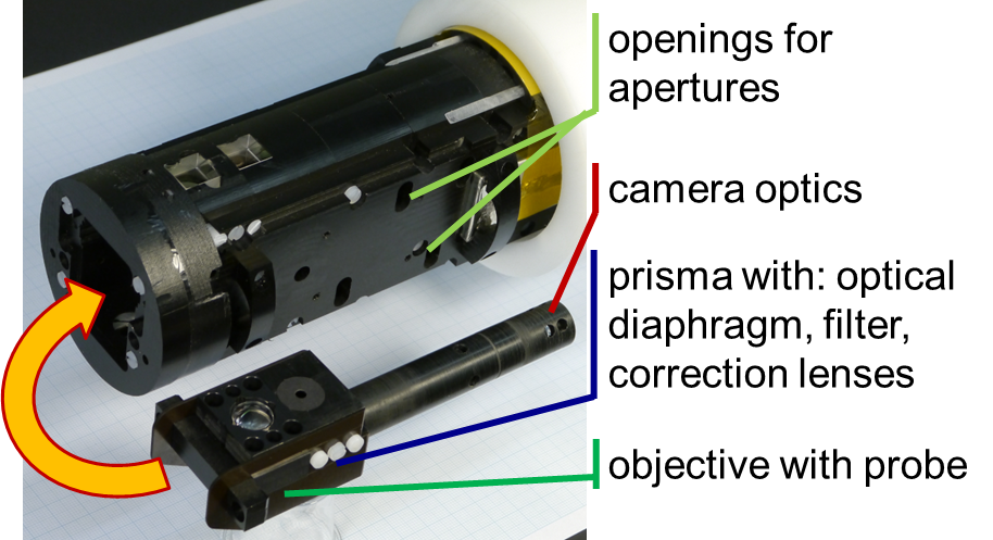
Front part of the microscope containing the optics. The objective can be take in and out from the microscope and can include a micro coil.
Magnetic Resonance (MR) microscopy is a method for high-resolution imaging resolving structures smaller than 100 µm. However, despite technical improvements of MRI, a lack of understanding remains regarding the effect of pathologies on a cellular level and their relations to MR contrast changes on a macroscopic level. The identification of specific microstructural elements is therefore done by light-microscopy based correlative histology techniques. In order to overcome these limitations, an Optical Microscope (OM) was build, which can be placed inside the bore of an ultra-high field (9.4 T) small animal MR scanner (Bruker Biospec 94/20) and allow to concurrently perform optical microscopy and MR imaging of the sample without compromises in the optical resolution. The OM has interchangeable optics with optical field of views of 0.5, 1 and 2 mm with sub-micrometer resolution. Optical microscopy can be performed with RGBW top and bottom, dark and bright field illumination. MR and optical microscopy can be performed simultaneously using a standard commercial quadrature coil or a Helmholtz-microcoil. The later was integrated into the objective where single-use sample containers can be placed for optical and MR microscopy. This research is performed in collaboration with the Laboratory for Microactuators, from the Department of Microsystems Engineering, IMTEK, University Freiburg The objective can be take in and out from the microscope and can include a microcoil.
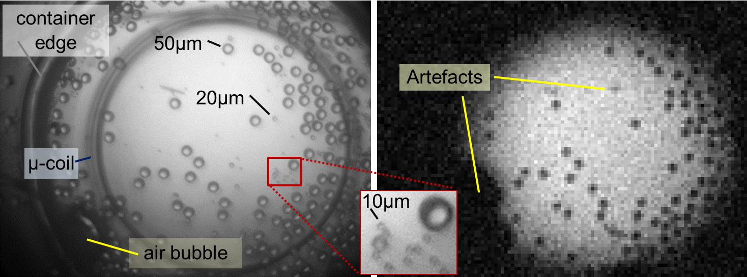
Glass beads observed with the OM (left) and with MR (right). The optical image was obtained by red bottom illumination and the MR image was acquired with a 2D GE sequence.

Dr. Sebastian Littin
Group Leader
Tel.: +49 761 270-93820
E-Mail: sebastian.littin@uniklinik-freiburg.de
University Medical Center Freiburg
Dept. of Radiology · Medical Physics
Killianstr. 5a
79106 Freiburg