Spatial Biology / Highly Multiplexed Microscopy: Akoya Phenocycler-Fusion (CODEX)
Spatial biology is an approach which looks at biomarker expression of single cells while maintaining their relative location within the tissue architecture.
The Akoya Phenocycler-Fusion is a two-part system consisting of an automated microscope and an automated fluidics system. The system is capable of highly multiplexed, high-resolution (20x, 40x) analysis of samples contained on single slides.
The first part of the system is the Fusion microscope, which is a multispectral, automated slide scanning system capable of examining up to 4 slides at a time using either fluorescence or brightfield imaging. The system has been optimized to work with the Akoya Motif OPAL dyes (6 biomarkers + DAPI), but is also compatible with most standard fluorescence dyes.
The second part of the system is the Phenocycler, also known as CODEX, which enables highly multiplexed, high resolution (20x, 40x) analysis of single slides. The sample slide is first labelled with antibodies which have been conjugated to oligonucleotide barcodes.
The Phenocycler itself is an automated fluidics control unit which enables successive rounds of staining using complementary barcoded oligonucleotides, labelled with fluorophores. The oligonucleotides bind to the already bound, barcoded antibodies, specifically labelling them. Recent reports have shown multiplexing or labelling of up to 100 markers.
For more information about the system or to give it a try, please contact the Lighthouse Core Facility.
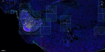
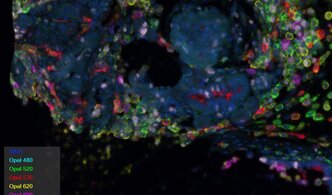
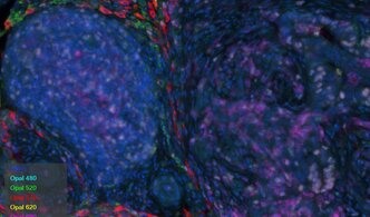
Examples of images acquired using the Lighthouse Akoya system can be found below:
More information about the Akoya Phenocycler-Fusion can also be found at the Akoya website.


