Microscopy
Long-Term Live Cell Imaging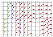
Sartorius Incucyte S3
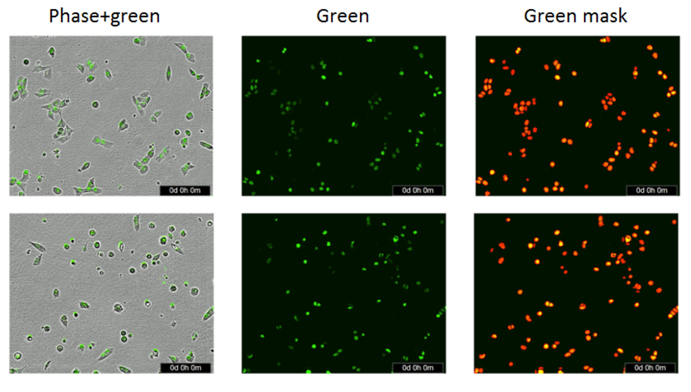
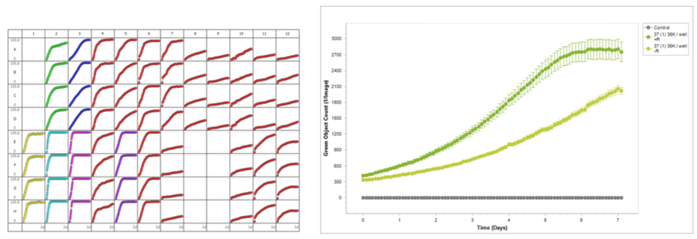
The Incucyte S3 is an automated microscope located within a standard cell culture incubator and is of particular interest for low resolution, long-term, live cell imaging. It allows the independent measurement and analysis of up to 6 plates or experiments at the same time. It is the method of choice for long-term observation of spheroid and organoid development. Available objectives include 4x, 10x and 20x. These objectives are compatible with most standard cell culture plate formats and plates of to 96- or 384-wells are also supported. For the list of currently supported substrates please see (link for download). Channels available for imaging and analysis are standard brightfield/phase contrast, as well as two channels for fluorescence. Details about fluorescence can be seen below:
|
Color |
Excitation |
Emission |
Possible Fluorochromes |
|
Green fluorescence |
440-480 nm |
504-544 nm |
GFP, YFP, CellTracker Green, CFSE, FITC |
|
Red fluorescence |
565-605 nm |
625-705 nm |
mRFP, mKate2, mCherry, CellTracker Red, Alexa Fluor 594 |
Note: The red fluorescence channel is suboptimal for the detection of fluorescence proteins such as td tomato and turbo RFP.

Plates and Flasks fitting into IncuCyte
The list for download contains the vessels which work with the IncuCyte. Note: Only Microwell Plate formats or chamber slides will fit into our system. No flasks.